Calcium »
PDB 6im3-6j43 »
6j42 »
Calcium in PDB 6j42: Crystal Structure of Wild Type Katb, A Manganese Catalase From Anabaena
Protein crystallography data
The structure of Crystal Structure of Wild Type Katb, A Manganese Catalase From Anabaena, PDB code: 6j42
was solved by
S.C.Bihani,
D.Chakravarty,
A.Ballal,
with X-Ray Crystallography technique. A brief refinement statistics is given in the table below:
| Resolution Low / High (Å) | 19.59 / 2.49 |
| Space group | P 41 21 2 |
| Cell size a, b, c (Å), α, β, γ (°) | 102.070, 102.070, 138.980, 90.00, 90.00, 90.00 |
| R / Rfree (%) | 21.7 / 26.1 |
Other elements in 6j42:
The structure of Crystal Structure of Wild Type Katb, A Manganese Catalase From Anabaena also contains other interesting chemical elements:
| Manganese | (Mn) | 6 atoms |
Calcium Binding Sites:
The binding sites of Calcium atom in the Crystal Structure of Wild Type Katb, A Manganese Catalase From Anabaena
(pdb code 6j42). This binding sites where shown within
5.0 Angstroms radius around Calcium atom.
In total 5 binding sites of Calcium where determined in the Crystal Structure of Wild Type Katb, A Manganese Catalase From Anabaena, PDB code: 6j42:
Jump to Calcium binding site number: 1; 2; 3; 4; 5;
In total 5 binding sites of Calcium where determined in the Crystal Structure of Wild Type Katb, A Manganese Catalase From Anabaena, PDB code: 6j42:
Jump to Calcium binding site number: 1; 2; 3; 4; 5;
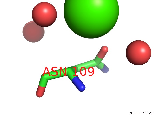
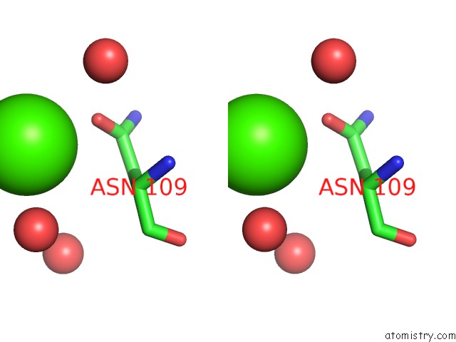
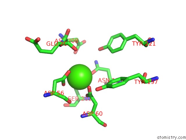
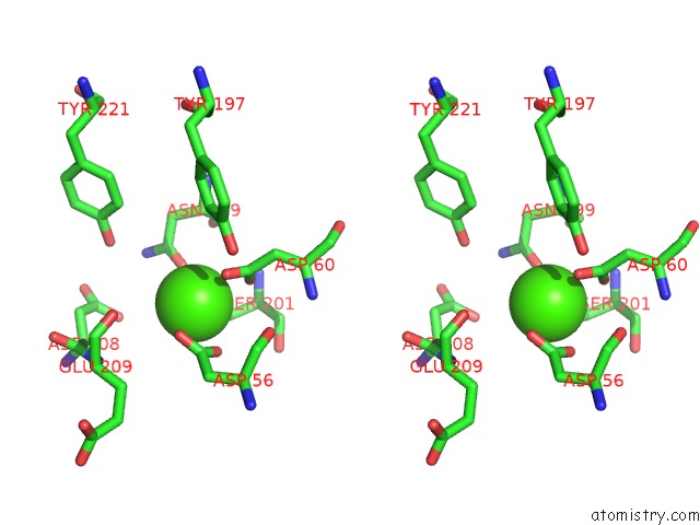
Calcium binding site 1 out of 5 in 6j42
Go back to
Calcium binding site 1 out
of 5 in the Crystal Structure of Wild Type Katb, A Manganese Catalase From Anabaena
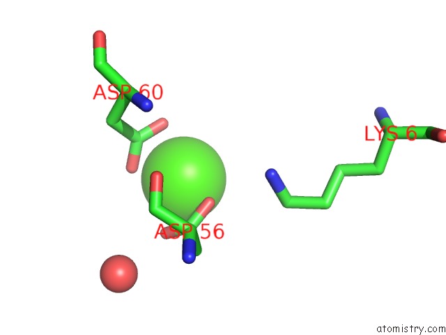
Mono view
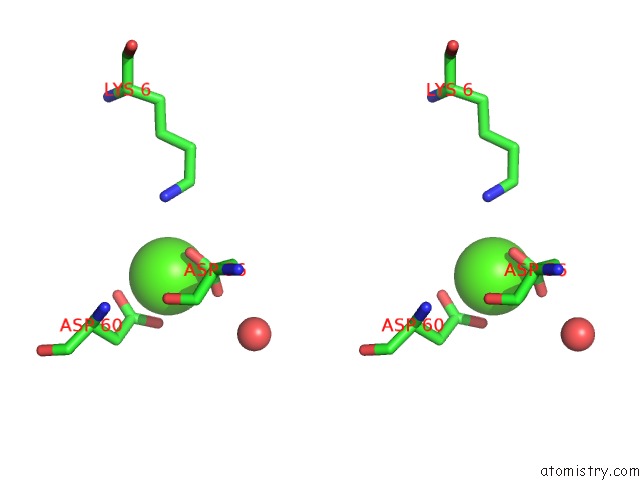
Stereo pair view
Mono view
Stereo pair view
A full contact list of Calcium with other atoms in the Ca binding
site number 1 of Crystal Structure of Wild Type Katb, A Manganese Catalase From Anabaena within 5.0Å range:
|
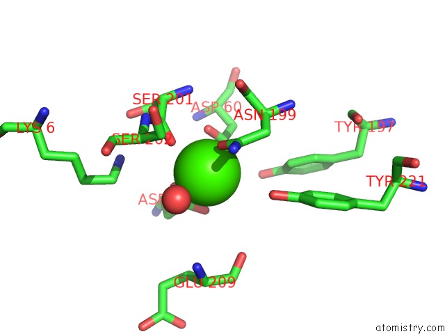
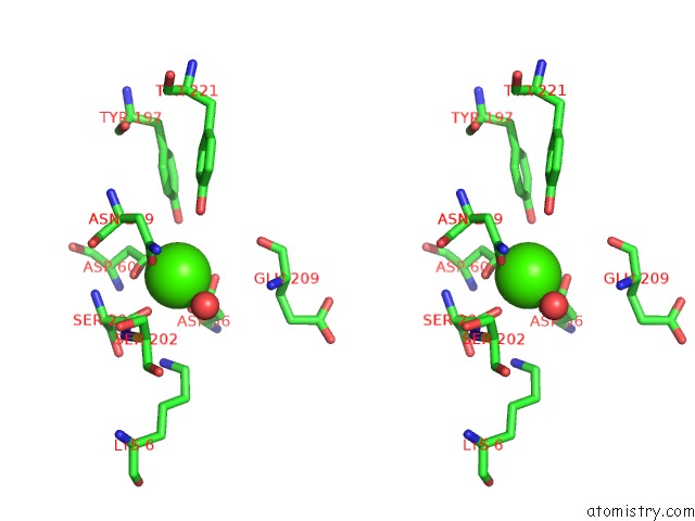
Calcium binding site 2 out of 5 in 6j42
Go back to
Calcium binding site 2 out
of 5 in the Crystal Structure of Wild Type Katb, A Manganese Catalase From Anabaena
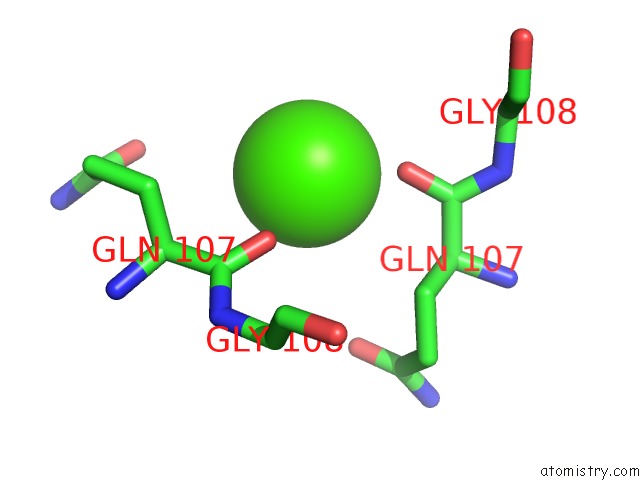
Mono view
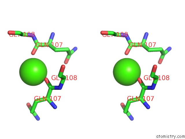
Stereo pair view
Mono view
Stereo pair view
A full contact list of Calcium with other atoms in the Ca binding
site number 2 of Crystal Structure of Wild Type Katb, A Manganese Catalase From Anabaena within 5.0Å range:
|
Calcium binding site 3 out of 5 in 6j42
Go back to
Calcium binding site 3 out
of 5 in the Crystal Structure of Wild Type Katb, A Manganese Catalase From Anabaena
Mono view
Stereo pair view
Mono view
Stereo pair view
A full contact list of Calcium with other atoms in the Ca binding
site number 3 of Crystal Structure of Wild Type Katb, A Manganese Catalase From Anabaena within 5.0Å range:
|
Calcium binding site 4 out of 5 in 6j42
Go back to
Calcium binding site 4 out
of 5 in the Crystal Structure of Wild Type Katb, A Manganese Catalase From Anabaena
Mono view
Stereo pair view
Mono view
Stereo pair view
A full contact list of Calcium with other atoms in the Ca binding
site number 4 of Crystal Structure of Wild Type Katb, A Manganese Catalase From Anabaena within 5.0Å range:
|
Calcium binding site 5 out of 5 in 6j42
Go back to
Calcium binding site 5 out
of 5 in the Crystal Structure of Wild Type Katb, A Manganese Catalase From Anabaena
Mono view
Stereo pair view
Mono view
Stereo pair view
A full contact list of Calcium with other atoms in the Ca binding
site number 5 of Crystal Structure of Wild Type Katb, A Manganese Catalase From Anabaena within 5.0Å range:
|
Reference:
D.Chakravarty,
S.C.Bihani,
M.Banerjee,
A.Ballal.
Molecular Basis of the Unique Role Played By An N-Terminal Non-Active Site Residue of Cyanobacterial Mn-Catalase in Structure-Function Maintenance To Be Published.
Page generated: Tue Jul 16 09:40:58 2024
Last articles
Zn in 9J0NZn in 9J0O
Zn in 9J0P
Zn in 9FJX
Zn in 9EKB
Zn in 9C0F
Zn in 9CAH
Zn in 9CH0
Zn in 9CH3
Zn in 9CH1