Calcium »
PDB 6j4h-6jlm »
6jfm »
Calcium in PDB 6jfm: MITOFUSIN2 (MFN2)_T111D
Protein crystallography data
The structure of MITOFUSIN2 (MFN2)_T111D, PDB code: 6jfm
was solved by
Y.J.Li,
Y.L.Cao,
J.X.Feng,
Y.B.Qi,
S.X.Meng,
J.F.Yang,
Y.T.Zhong,
S.S.Kang,
X.X.Chen,
L.Lan,
L.Luo,
B.Yu,
S.D.Chen,
D.C.Chan,
J.J.Hu,
S.Gao,
with X-Ray Crystallography technique. A brief refinement statistics is given in the table below:
| Resolution Low / High (Å) | 31.64 / 2.09 |
| Space group | P 1 21 1 |
| Cell size a, b, c (Å), α, β, γ (°) | 44.437, 126.563, 79.972, 90.00, 102.46, 90.00 |
| R / Rfree (%) | 17.6 / 22.9 |
Calcium Binding Sites:
The binding sites of Calcium atom in the MITOFUSIN2 (MFN2)_T111D
(pdb code 6jfm). This binding sites where shown within
5.0 Angstroms radius around Calcium atom.
In total 3 binding sites of Calcium where determined in the MITOFUSIN2 (MFN2)_T111D, PDB code: 6jfm:
Jump to Calcium binding site number: 1; 2; 3;
In total 3 binding sites of Calcium where determined in the MITOFUSIN2 (MFN2)_T111D, PDB code: 6jfm:
Jump to Calcium binding site number: 1; 2; 3;
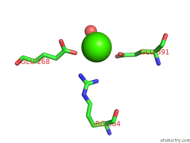
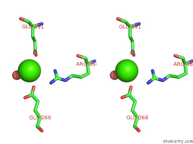
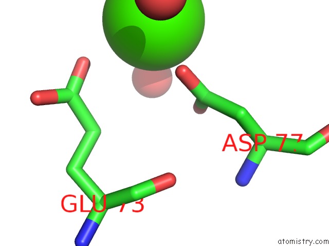
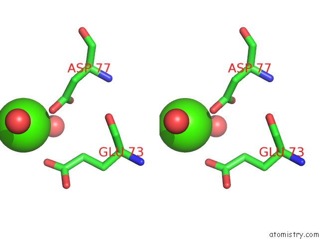
Calcium binding site 1 out of 3 in 6jfm
Go back to
Calcium binding site 1 out
of 3 in the MITOFUSIN2 (MFN2)_T111D
Mono view
Stereo pair view
Mono view
Stereo pair view
A full contact list of Calcium with other atoms in the Ca binding
site number 1 of MITOFUSIN2 (MFN2)_T111D within 5.0Å range:
|
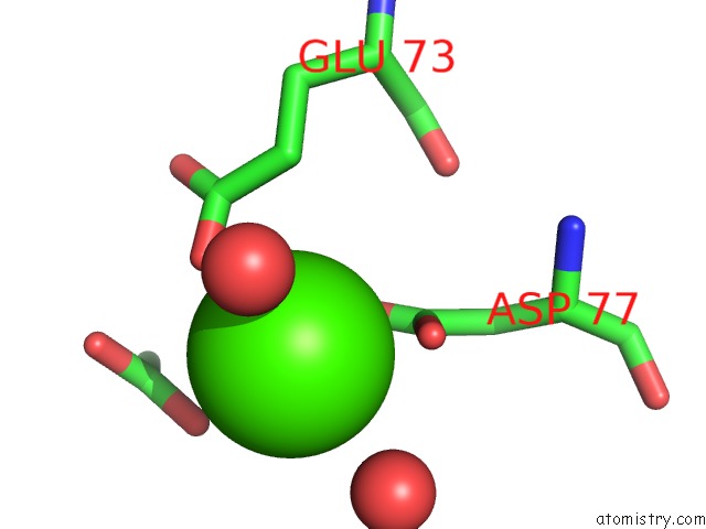
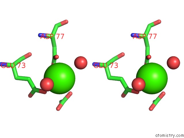
Calcium binding site 2 out of 3 in 6jfm
Go back to
Calcium binding site 2 out
of 3 in the MITOFUSIN2 (MFN2)_T111D
Mono view
Stereo pair view
Mono view
Stereo pair view
A full contact list of Calcium with other atoms in the Ca binding
site number 2 of MITOFUSIN2 (MFN2)_T111D within 5.0Å range:
|
Calcium binding site 3 out of 3 in 6jfm
Go back to
Calcium binding site 3 out
of 3 in the MITOFUSIN2 (MFN2)_T111D
Mono view
Stereo pair view
Mono view
Stereo pair view
A full contact list of Calcium with other atoms in the Ca binding
site number 3 of MITOFUSIN2 (MFN2)_T111D within 5.0Å range:
|
Reference:
Y.J.Li,
Y.L.Cao,
J.X.Feng,
Y.Qi,
S.Meng,
J.F.Yang,
Y.T.Zhong,
S.Kang,
X.Chen,
L.Lan,
L.Luo,
B.Yu,
S.Chen,
D.C.Chan,
J.Hu,
S.Gao.
Structural Insights of Human Mitofusin-2 Into Mitochondrial Fusion and CMT2A Onset. Nat Commun V. 10 4914 2019.
ISSN: ESSN 2041-1723
PubMed: 31664033
DOI: 10.1038/S41467-019-12912-0
Page generated: Wed Jul 9 15:06:52 2025
ISSN: ESSN 2041-1723
PubMed: 31664033
DOI: 10.1038/S41467-019-12912-0
Last articles
Cl in 5L3HCl in 5L3I
Cl in 5L2O
Cl in 5L3X
Cl in 5L39
Cl in 5L38
Cl in 5L35
Cl in 5L2U
Cl in 5L2N
Cl in 5L1E