Calcium »
PDB 6j4h-6jlm »
6jl9 »
Calcium in PDB 6jl9: Crystal Structure of A Frog Ependymin Related Protein
Protein crystallography data
The structure of Crystal Structure of A Frog Ependymin Related Protein, PDB code: 6jl9
was solved by
S.Y.Park,
with X-Ray Crystallography technique. A brief refinement statistics is given in the table below:
| Resolution Low / High (Å) | 53.01 / 2.00 |
| Space group | P 65 2 2 |
| Cell size a, b, c (Å), α, β, γ (°) | 61.208, 61.208, 236.196, 90.00, 90.00, 120.00 |
| R / Rfree (%) | 19.4 / 27.8 |
Calcium Binding Sites:
The binding sites of Calcium atom in the Crystal Structure of A Frog Ependymin Related Protein
(pdb code 6jl9). This binding sites where shown within
5.0 Angstroms radius around Calcium atom.
In total only one binding site of Calcium was determined in the Crystal Structure of A Frog Ependymin Related Protein, PDB code: 6jl9:
In total only one binding site of Calcium was determined in the Crystal Structure of A Frog Ependymin Related Protein, PDB code: 6jl9:
Calcium binding site 1 out of 1 in 6jl9
Go back to
Calcium binding site 1 out
of 1 in the Crystal Structure of A Frog Ependymin Related Protein
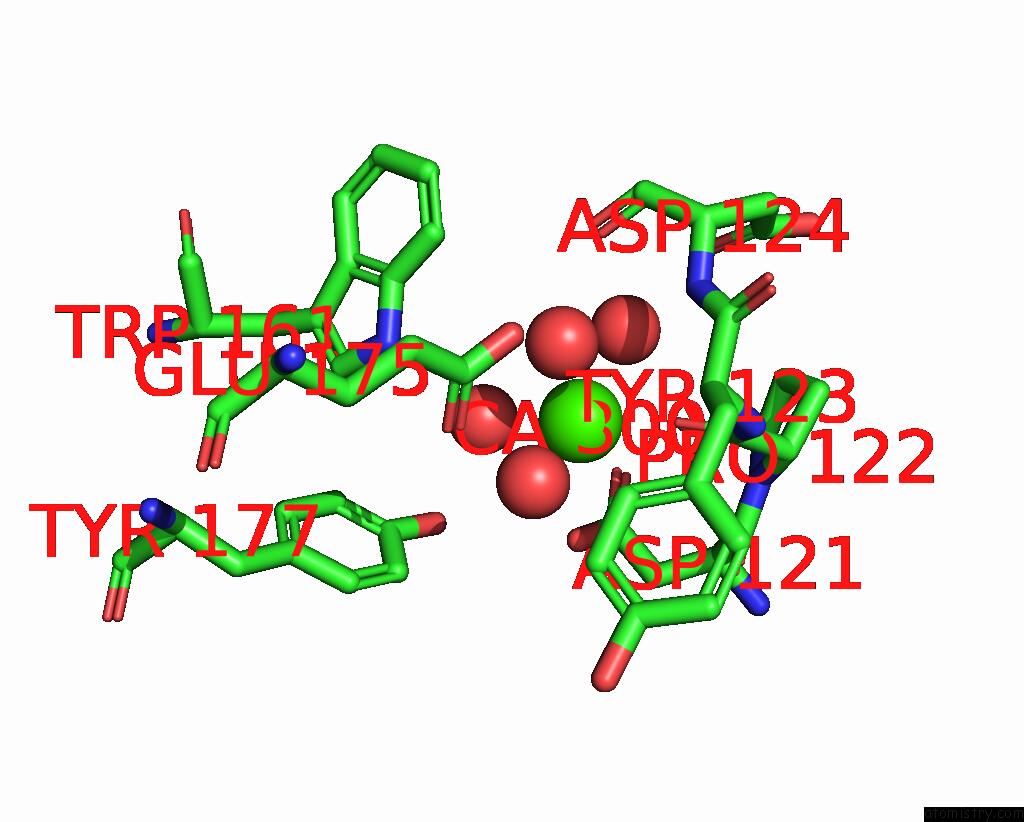
Mono view
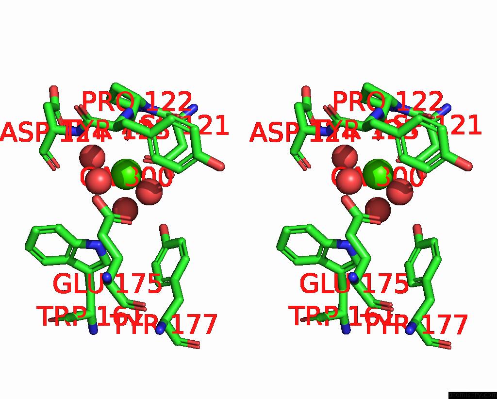
Stereo pair view
Mono view
Stereo pair view
A full contact list of Calcium with other atoms in the Ca binding
site number 1 of Crystal Structure of A Frog Ependymin Related Protein within 5.0Å range:
|
Reference:
J.K.Park,
K.Y.Kim,
Y.W.Sim,
Y.I.Kim,
J.K.Kim,
C.Lee,
J.Han,
C.U.Kim,
J.E.Lee,
S.Park.
Structures of Three Ependymin-Related Proteins Suggest Their Function As A Hydrophobic Molecule Binder. Iucrj V. 6 729 2019.
ISSN: ESSN 2052-2525
PubMed: 31316816
DOI: 10.1107/S2052252519007668
Page generated: Tue Jul 16 09:51:45 2024
ISSN: ESSN 2052-2525
PubMed: 31316816
DOI: 10.1107/S2052252519007668
Last articles
Zn in 9J0NZn in 9J0O
Zn in 9J0P
Zn in 9FJX
Zn in 9EKB
Zn in 9C0F
Zn in 9CAH
Zn in 9CH0
Zn in 9CH3
Zn in 9CH1