Calcium »
PDB 6jln-6k2p »
6jt5 »
Calcium in PDB 6jt5: Crystal Structure of Pqq Doamin of Pyranose Dehydrogenase From Coprinopsis Cinerea: Apo-From
Protein crystallography data
The structure of Crystal Structure of Pqq Doamin of Pyranose Dehydrogenase From Coprinopsis Cinerea: Apo-From, PDB code: 6jt5
was solved by
K.Takeda,
T.Ishida,
M.Yoshida,
M.Samejima,
H.Ohno,
K.Igarashi,
N.Nakamura,
with X-Ray Crystallography technique. A brief refinement statistics is given in the table below:
| Resolution Low / High (Å) | 19.75 / 1.50 |
| Space group | P 1 21 1 |
| Cell size a, b, c (Å), α, β, γ (°) | 62.298, 47.851, 69.375, 90.00, 115.55, 90.00 |
| R / Rfree (%) | 10.5 / 13.4 |
Calcium Binding Sites:
The binding sites of Calcium atom in the Crystal Structure of Pqq Doamin of Pyranose Dehydrogenase From Coprinopsis Cinerea: Apo-From
(pdb code 6jt5). This binding sites where shown within
5.0 Angstroms radius around Calcium atom.
In total only one binding site of Calcium was determined in the Crystal Structure of Pqq Doamin of Pyranose Dehydrogenase From Coprinopsis Cinerea: Apo-From, PDB code: 6jt5:
In total only one binding site of Calcium was determined in the Crystal Structure of Pqq Doamin of Pyranose Dehydrogenase From Coprinopsis Cinerea: Apo-From, PDB code: 6jt5:
Calcium binding site 1 out of 1 in 6jt5
Go back to
Calcium binding site 1 out
of 1 in the Crystal Structure of Pqq Doamin of Pyranose Dehydrogenase From Coprinopsis Cinerea: Apo-From
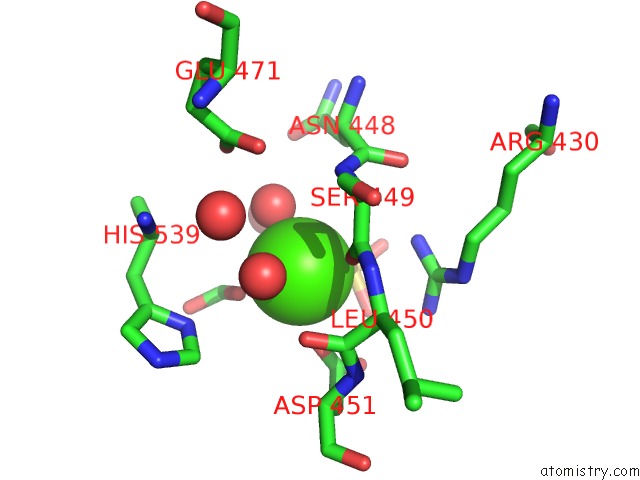
Mono view
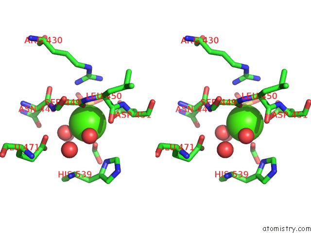
Stereo pair view
Mono view
Stereo pair view
A full contact list of Calcium with other atoms in the Ca binding
site number 1 of Crystal Structure of Pqq Doamin of Pyranose Dehydrogenase From Coprinopsis Cinerea: Apo-From within 5.0Å range:
|
Reference:
K.Takeda,
T.Ishida,
M.Yoshida,
M.Samejima,
H.Ohno,
K.Igarashi,
N.Nakamura.
The Crystal Structure of the Catalytic Domain and the Cytochromebdomain in A Eukaryotic Pqq-Dependent Dehydrogenase. Appl.Environ.Microbiol. 2019.
ISSN: ESSN 1098-5336
PubMed: 31604769
DOI: 10.1128/AEM.01692-19
Page generated: Tue Jul 16 10:06:32 2024
ISSN: ESSN 1098-5336
PubMed: 31604769
DOI: 10.1128/AEM.01692-19
Last articles
Zn in 9J0NZn in 9J0O
Zn in 9J0P
Zn in 9FJX
Zn in 9EKB
Zn in 9C0F
Zn in 9CAH
Zn in 9CH0
Zn in 9CH3
Zn in 9CH1