Calcium »
PDB 6jlm-6k22 »
6k0p »
Calcium in PDB 6k0p: Catalytic Domain of GH87 Alpha-1,3-Glucanase D1045A in Complex with Nigerose
Protein crystallography data
The structure of Catalytic Domain of GH87 Alpha-1,3-Glucanase D1045A in Complex with Nigerose, PDB code: 6k0p
was solved by
T.Itoh,
R.Intuy,
W.Suyotha,
J.Hayashi,
S.Yano,
K.Makabe,
M.Wakayama,
T.Hibi,
with X-Ray Crystallography technique. A brief refinement statistics is given in the table below:
| Resolution Low / High (Å) | 36.76 / 1.42 |
| Space group | P 43 21 2 |
| Cell size a, b, c (Å), α, β, γ (°) | 132.525, 132.525, 76.147, 90.00, 90.00, 90.00 |
| R / Rfree (%) | 15.6 / 17 |
Calcium Binding Sites:
The binding sites of Calcium atom in the Catalytic Domain of GH87 Alpha-1,3-Glucanase D1045A in Complex with Nigerose
(pdb code 6k0p). This binding sites where shown within
5.0 Angstroms radius around Calcium atom.
In total only one binding site of Calcium was determined in the Catalytic Domain of GH87 Alpha-1,3-Glucanase D1045A in Complex with Nigerose, PDB code: 6k0p:
In total only one binding site of Calcium was determined in the Catalytic Domain of GH87 Alpha-1,3-Glucanase D1045A in Complex with Nigerose, PDB code: 6k0p:
Calcium binding site 1 out of 1 in 6k0p
Go back to
Calcium binding site 1 out
of 1 in the Catalytic Domain of GH87 Alpha-1,3-Glucanase D1045A in Complex with Nigerose
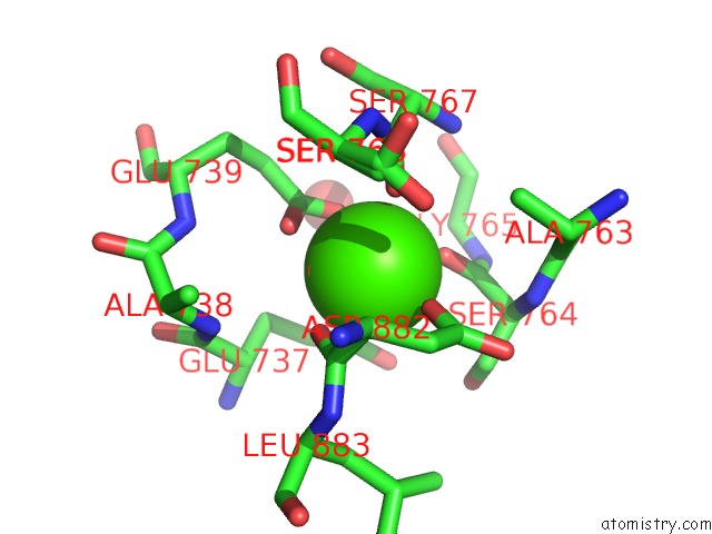
Mono view
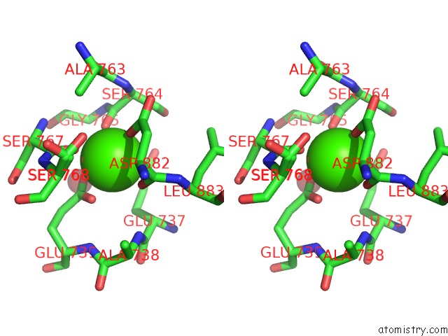
Stereo pair view
Mono view
Stereo pair view
A full contact list of Calcium with other atoms in the Ca binding
site number 1 of Catalytic Domain of GH87 Alpha-1,3-Glucanase D1045A in Complex with Nigerose within 5.0Å range:
|
Reference:
T.Itoh,
R.Intuy,
W.Suyotha,
J.Hayashi,
S.Yano,
K.Makabe,
M.Wakayama,
T.Hibi.
28STRUCTURAL Insights Into Substrate Recognition and Catalysis By Glycoside Hydrolase Family 87 Alpha-1,3-Glucanase From Paenibacillus Glycanilyticus FH11. Febs J. 2019.
ISSN: ISSN 1742-464X
PubMed: 31788942
DOI: 10.1111/FEBS.15161
Page generated: Tue Jul 16 10:10:54 2024
ISSN: ISSN 1742-464X
PubMed: 31788942
DOI: 10.1111/FEBS.15161
Last articles
Zn in 9JYWZn in 9IR4
Zn in 9IR3
Zn in 9GMX
Zn in 9GMW
Zn in 9JEJ
Zn in 9ERF
Zn in 9ERE
Zn in 9EGV
Zn in 9EGW