Calcium »
PDB 6kg9-6kzk »
6kgb »
Calcium in PDB 6kgb: Crystal Structure of E61K Mutated Transthyretin
Protein crystallography data
The structure of Crystal Structure of E61K Mutated Transthyretin, PDB code: 6kgb
was solved by
T.Yokoyama,
M.Mizuguchi,
with X-Ray Crystallography technique. A brief refinement statistics is given in the table below:
| Resolution Low / High (Å) | 42.80 / 1.30 |
| Space group | P 21 21 2 |
| Cell size a, b, c (Å), α, β, γ (°) | 42.538, 85.597, 64.419, 90.00, 90.00, 90.00 |
| R / Rfree (%) | 16.1 / 17.7 |
Calcium Binding Sites:
The binding sites of Calcium atom in the Crystal Structure of E61K Mutated Transthyretin
(pdb code 6kgb). This binding sites where shown within
5.0 Angstroms radius around Calcium atom.
In total only one binding site of Calcium was determined in the Crystal Structure of E61K Mutated Transthyretin, PDB code: 6kgb:
In total only one binding site of Calcium was determined in the Crystal Structure of E61K Mutated Transthyretin, PDB code: 6kgb:
Calcium binding site 1 out of 1 in 6kgb
Go back to
Calcium binding site 1 out
of 1 in the Crystal Structure of E61K Mutated Transthyretin
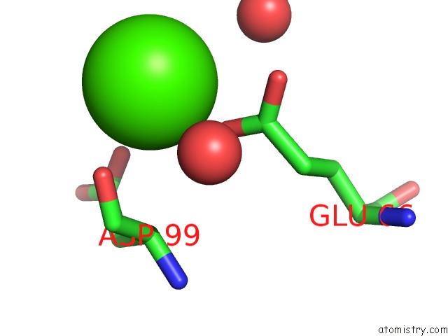
Mono view
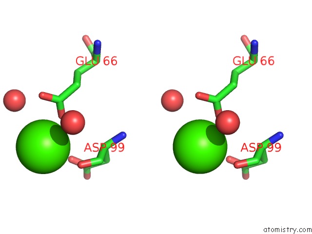
Stereo pair view
Mono view
Stereo pair view
A full contact list of Calcium with other atoms in the Ca binding
site number 1 of Crystal Structure of E61K Mutated Transthyretin within 5.0Å range:
|
Reference:
T.Murakami,
T.Yokoyama,
M.Mizuguchi,
S.Tone,
S.Takaku,
K.Sango,
H.Nishimura,
K.Watabe,
Y.Sunada.
A Low Amyloidogenic E61K Transthyretin Mutation May Cause Familial Amyloid Polyneuropathy. J.Neurochem. 2020.
ISSN: ESSN 1471-4159
PubMed: 32852783
DOI: 10.1111/JNC.15162
Page generated: Tue Jul 16 10:28:16 2024
ISSN: ESSN 1471-4159
PubMed: 32852783
DOI: 10.1111/JNC.15162
Last articles
Zn in 9J0NZn in 9J0O
Zn in 9J0P
Zn in 9FJX
Zn in 9EKB
Zn in 9C0F
Zn in 9CAH
Zn in 9CH0
Zn in 9CH3
Zn in 9CH1