Calcium »
PDB 6kg9-6kzk »
6kqu »
Calcium in PDB 6kqu: Crystal Structure of Phospholipase A2
Enzymatic activity of Crystal Structure of Phospholipase A2
All present enzymatic activity of Crystal Structure of Phospholipase A2:
3.1.1.4;
3.1.1.4;
Protein crystallography data
The structure of Crystal Structure of Phospholipase A2, PDB code: 6kqu
was solved by
S.Hou,
T.Xu,
J.Liu,
with X-Ray Crystallography technique. A brief refinement statistics is given in the table below:
| Resolution Low / High (Å) | 18.80 / 2.00 |
| Space group | P 21 2 2 |
| Cell size a, b, c (Å), α, β, γ (°) | 48.839, 61.117, 63.404, 90.00, 90.00, 90.00 |
| R / Rfree (%) | 19.7 / 23.9 |
Other elements in 6kqu:
The structure of Crystal Structure of Phospholipase A2 also contains other interesting chemical elements:
| Chlorine | (Cl) | 6 atoms |
| Sodium | (Na) | 1 atom |
Calcium Binding Sites:
The binding sites of Calcium atom in the Crystal Structure of Phospholipase A2
(pdb code 6kqu). This binding sites where shown within
5.0 Angstroms radius around Calcium atom.
In total only one binding site of Calcium was determined in the Crystal Structure of Phospholipase A2, PDB code: 6kqu:
In total only one binding site of Calcium was determined in the Crystal Structure of Phospholipase A2, PDB code: 6kqu:
Calcium binding site 1 out of 1 in 6kqu
Go back to
Calcium binding site 1 out
of 1 in the Crystal Structure of Phospholipase A2
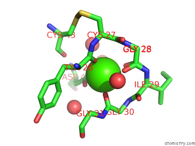
Mono view
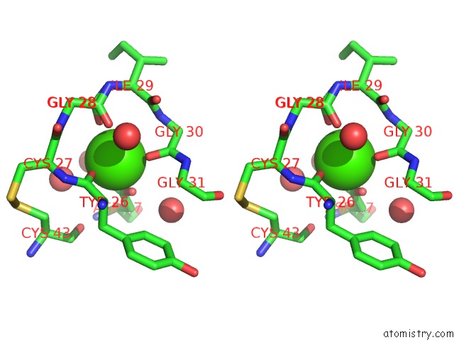
Stereo pair view
Mono view
Stereo pair view
A full contact list of Calcium with other atoms in the Ca binding
site number 1 of Crystal Structure of Phospholipase A2 within 5.0Å range:
|
Reference:
S.Hou,
Y.Zhang,
J.Xu,
J.Bai,
J.Liu,
J.Xie,
T.Xu.
Residue ASN21 Acts As A Switch For Calcium Binding to Modulate the Enzymatic Activity of Human Phospholipase A2 Group Iie. Biochimie V. 176 117 2020.
ISSN: ISSN 0300-9084
PubMed: 32659444
DOI: 10.1016/J.BIOCHI.2020.07.003
Page generated: Tue Jul 16 10:37:10 2024
ISSN: ISSN 0300-9084
PubMed: 32659444
DOI: 10.1016/J.BIOCHI.2020.07.003
Last articles
Zn in 9J0NZn in 9J0O
Zn in 9J0P
Zn in 9FJX
Zn in 9EKB
Zn in 9C0F
Zn in 9CAH
Zn in 9CH0
Zn in 9CH3
Zn in 9CH1