Calcium »
PDB 6m4s-6mrl »
6m4s »
Calcium in PDB 6m4s: Crystal Structure Analysis of the Cytochrome P450 Cyp-SB21
Protein crystallography data
The structure of Crystal Structure Analysis of the Cytochrome P450 Cyp-SB21, PDB code: 6m4s
was solved by
F.W.Li,
S.Y.Li,
with X-Ray Crystallography technique. A brief refinement statistics is given in the table below:
| Resolution Low / High (Å) | 50.00 / 1.85 |
| Space group | P 1 21 1 |
| Cell size a, b, c (Å), α, β, γ (°) | 41.636, 91.092, 52.975, 90, 94.65, 90 |
| R / Rfree (%) | 15.1 / 19.7 |
Other elements in 6m4s:
The structure of Crystal Structure Analysis of the Cytochrome P450 Cyp-SB21 also contains other interesting chemical elements:
| Iron | (Fe) | 1 atom |
Calcium Binding Sites:
The binding sites of Calcium atom in the Crystal Structure Analysis of the Cytochrome P450 Cyp-SB21
(pdb code 6m4s). This binding sites where shown within
5.0 Angstroms radius around Calcium atom.
In total only one binding site of Calcium was determined in the Crystal Structure Analysis of the Cytochrome P450 Cyp-SB21, PDB code: 6m4s:
In total only one binding site of Calcium was determined in the Crystal Structure Analysis of the Cytochrome P450 Cyp-SB21, PDB code: 6m4s:
Calcium binding site 1 out of 1 in 6m4s
Go back to
Calcium binding site 1 out
of 1 in the Crystal Structure Analysis of the Cytochrome P450 Cyp-SB21
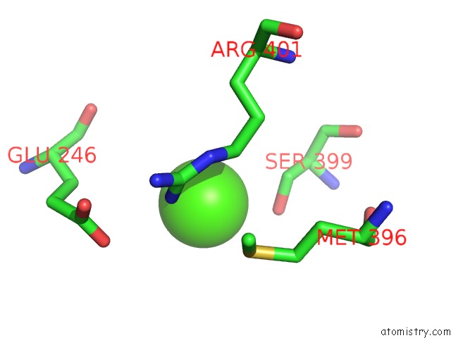
Mono view
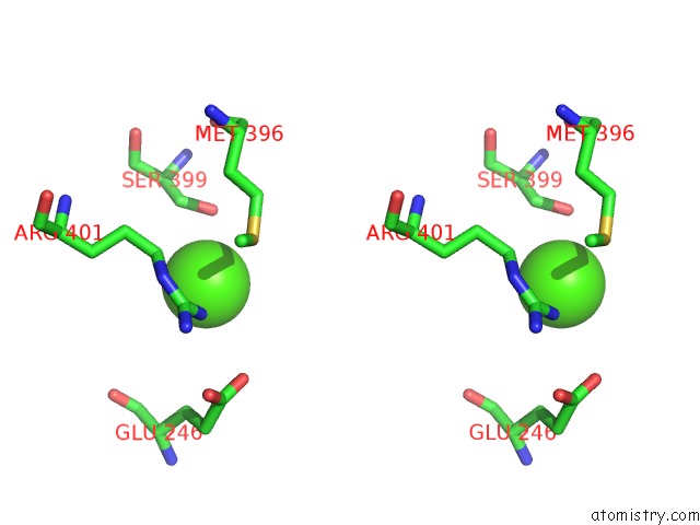
Stereo pair view
Mono view
Stereo pair view
A full contact list of Calcium with other atoms in the Ca binding
site number 1 of Crystal Structure Analysis of the Cytochrome P450 Cyp-SB21 within 5.0Å range:
|
Reference:
F.Li,
L.Ma,
X.Zhang,
J.Chen,
F.Qi,
Y.Huang,
Z.Qu,
L.Yao,
W.Zhang,
E.S.Kim,
S.Li.
Structure-Guided Manipulation of the Regioselectivity of the Cyclosporine A Hydroxylase Cyp-SB21 From Sebekia Benihana . Synth Syst Biotechnol V. 5 236 2020.
ISSN: ISSN 2405-805X
PubMed: 32775708
DOI: 10.1016/J.SYNBIO.2020.07.004
Page generated: Tue Jul 16 11:14:37 2024
ISSN: ISSN 2405-805X
PubMed: 32775708
DOI: 10.1016/J.SYNBIO.2020.07.004
Last articles
Zn in 9J0NZn in 9J0O
Zn in 9J0P
Zn in 9FJX
Zn in 9EKB
Zn in 9C0F
Zn in 9CAH
Zn in 9CH0
Zn in 9CH3
Zn in 9CH1