Calcium »
PDB 6m5a-6mrm »
6mf0 »
Calcium in PDB 6mf0: Crystal Structure Determination of Human/Porcine Chimera Coagulation Factor VIII
Protein crystallography data
The structure of Crystal Structure Determination of Human/Porcine Chimera Coagulation Factor VIII, PDB code: 6mf0
was solved by
I.W.Smith,
P.C.Spiegel,
with X-Ray Crystallography technique. A brief refinement statistics is given in the table below:
| Resolution Low / High (Å) | 49.03 / 3.20 |
| Space group | P 1 21 1 |
| Cell size a, b, c (Å), α, β, γ (°) | 72.005, 135.863, 196.107, 90.00, 90.15, 90.00 |
| R / Rfree (%) | 20.6 / 28.7 |
Other elements in 6mf0:
The structure of Crystal Structure Determination of Human/Porcine Chimera Coagulation Factor VIII also contains other interesting chemical elements:
| Copper | (Cu) | 2 atoms |
| Zinc | (Zn) | 2 atoms |
Calcium Binding Sites:
The binding sites of Calcium atom in the Crystal Structure Determination of Human/Porcine Chimera Coagulation Factor VIII
(pdb code 6mf0). This binding sites where shown within
5.0 Angstroms radius around Calcium atom.
In total 2 binding sites of Calcium where determined in the Crystal Structure Determination of Human/Porcine Chimera Coagulation Factor VIII, PDB code: 6mf0:
Jump to Calcium binding site number: 1; 2;
In total 2 binding sites of Calcium where determined in the Crystal Structure Determination of Human/Porcine Chimera Coagulation Factor VIII, PDB code: 6mf0:
Jump to Calcium binding site number: 1; 2;
Calcium binding site 1 out of 2 in 6mf0
Go back to
Calcium binding site 1 out
of 2 in the Crystal Structure Determination of Human/Porcine Chimera Coagulation Factor VIII
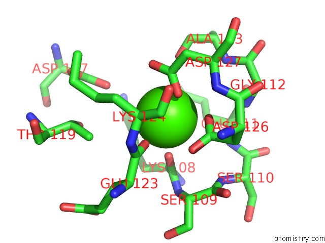
Mono view
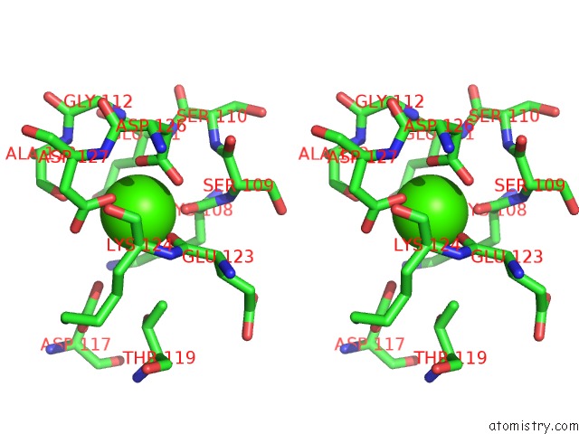
Stereo pair view
Mono view
Stereo pair view
A full contact list of Calcium with other atoms in the Ca binding
site number 1 of Crystal Structure Determination of Human/Porcine Chimera Coagulation Factor VIII within 5.0Å range:
|
Calcium binding site 2 out of 2 in 6mf0
Go back to
Calcium binding site 2 out
of 2 in the Crystal Structure Determination of Human/Porcine Chimera Coagulation Factor VIII
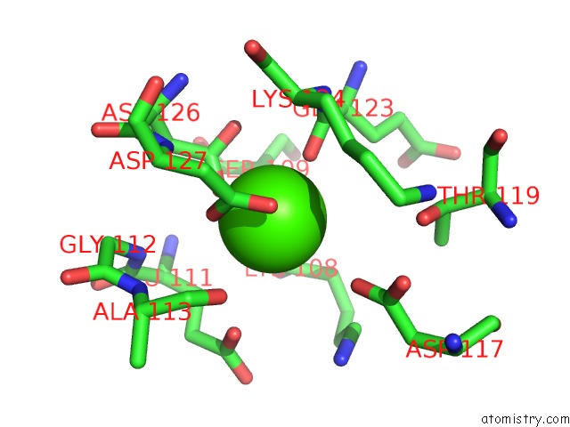
Mono view
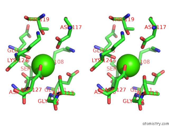
Stereo pair view
Mono view
Stereo pair view
A full contact list of Calcium with other atoms in the Ca binding
site number 2 of Crystal Structure Determination of Human/Porcine Chimera Coagulation Factor VIII within 5.0Å range:
|
Reference:
I.W.Smith,
A.E.D'aquino,
C.W.Coyle,
A.Fedanov,
E.T.Parker,
G.Denning,
H.T.Spencer,
P.Lollar,
C.B.Doering,
P.C.Spiegel Jr..
The 3.2 Angstrom Structure of A Bioengineered Variant of Blood Coagulation Factor VIII Indicates Two Conformations of the C2 Domain. J.Thromb.Haemost. 2019.
ISSN: ESSN 1538-7836
PubMed: 31454152
DOI: 10.1111/JTH.14621
Page generated: Wed Jul 9 16:05:18 2025
ISSN: ESSN 1538-7836
PubMed: 31454152
DOI: 10.1111/JTH.14621
Last articles
F in 7MYYF in 7N13
F in 7MYU
F in 7MYR
F in 7MYO
F in 7MXN
F in 7MXG
F in 7MXH
F in 7MX7
F in 7MVS