Calcium »
PDB 6m5a-6mrm »
6mga »
Calcium in PDB 6mga: Crystal Structure of Human Protocadherin-1 EC1-4 with Glycosylation
Protein crystallography data
The structure of Crystal Structure of Human Protocadherin-1 EC1-4 with Glycosylation, PDB code: 6mga
was solved by
D.Modak,
M.Sotomayor,
with X-Ray Crystallography technique. A brief refinement statistics is given in the table below:
| Resolution Low / High (Å) | 48.53 / 3.15 |
| Space group | P 61 2 2 |
| Cell size a, b, c (Å), α, β, γ (°) | 147.227, 147.227, 149.366, 90.00, 90.00, 120.00 |
| R / Rfree (%) | 21.5 / 26.3 |
Calcium Binding Sites:
The binding sites of Calcium atom in the Crystal Structure of Human Protocadherin-1 EC1-4 with Glycosylation
(pdb code 6mga). This binding sites where shown within
5.0 Angstroms radius around Calcium atom.
In total 9 binding sites of Calcium where determined in the Crystal Structure of Human Protocadherin-1 EC1-4 with Glycosylation, PDB code: 6mga:
Jump to Calcium binding site number: 1; 2; 3; 4; 5; 6; 7; 8; 9;
In total 9 binding sites of Calcium where determined in the Crystal Structure of Human Protocadherin-1 EC1-4 with Glycosylation, PDB code: 6mga:
Jump to Calcium binding site number: 1; 2; 3; 4; 5; 6; 7; 8; 9;
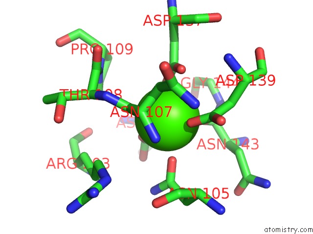
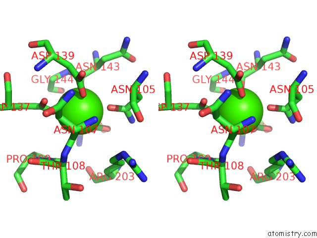
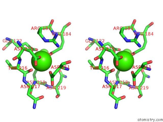
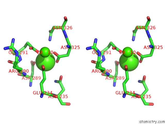
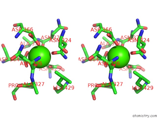
Calcium binding site 1 out of 9 in 6mga
Go back to
Calcium binding site 1 out
of 9 in the Crystal Structure of Human Protocadherin-1 EC1-4 with Glycosylation
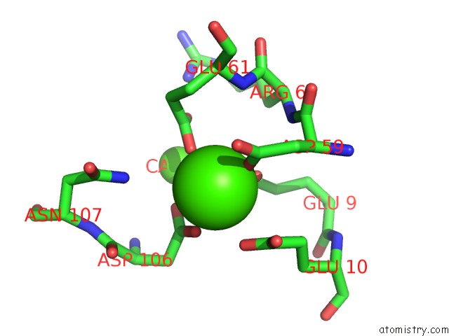
Mono view
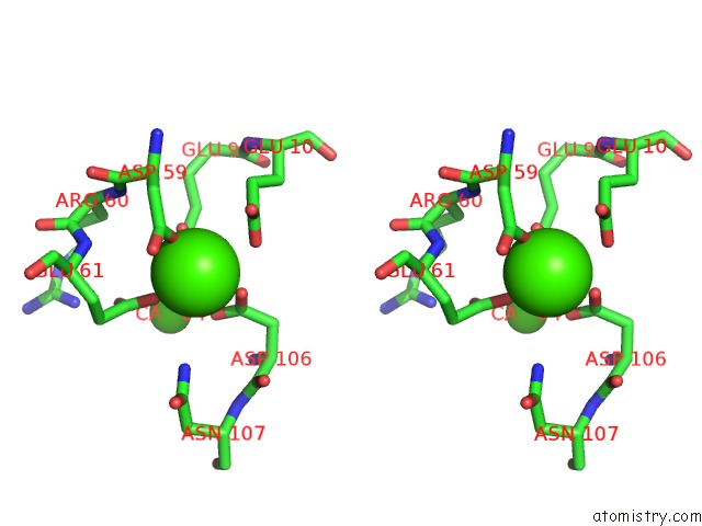
Stereo pair view
Mono view
Stereo pair view
A full contact list of Calcium with other atoms in the Ca binding
site number 1 of Crystal Structure of Human Protocadherin-1 EC1-4 with Glycosylation within 5.0Å range:
|
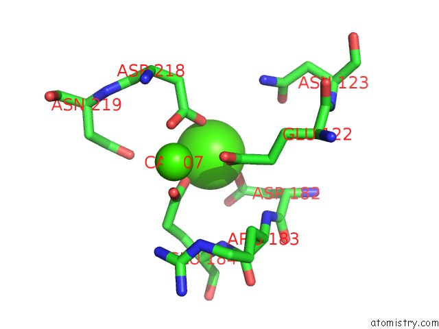
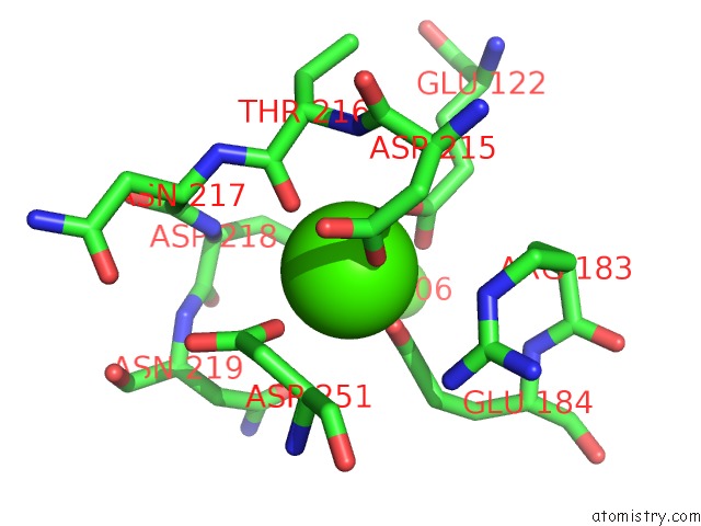
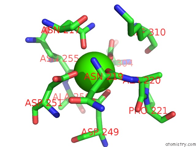
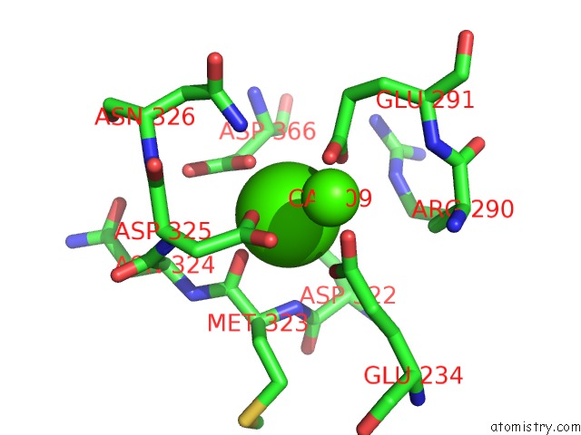
Calcium binding site 2 out of 9 in 6mga
Go back to
Calcium binding site 2 out
of 9 in the Crystal Structure of Human Protocadherin-1 EC1-4 with Glycosylation
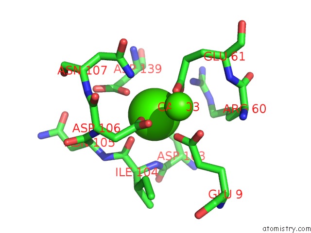
Mono view
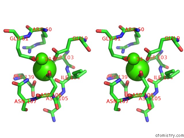
Stereo pair view
Mono view
Stereo pair view
A full contact list of Calcium with other atoms in the Ca binding
site number 2 of Crystal Structure of Human Protocadherin-1 EC1-4 with Glycosylation within 5.0Å range:
|
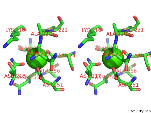
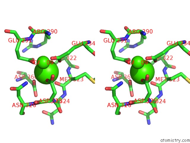
Calcium binding site 3 out of 9 in 6mga
Go back to
Calcium binding site 3 out
of 9 in the Crystal Structure of Human Protocadherin-1 EC1-4 with Glycosylation
Mono view
Stereo pair view
Mono view
Stereo pair view
A full contact list of Calcium with other atoms in the Ca binding
site number 3 of Crystal Structure of Human Protocadherin-1 EC1-4 with Glycosylation within 5.0Å range:
|
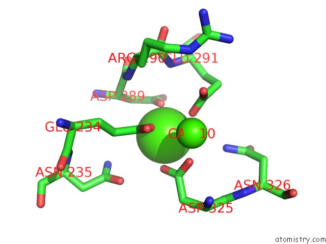
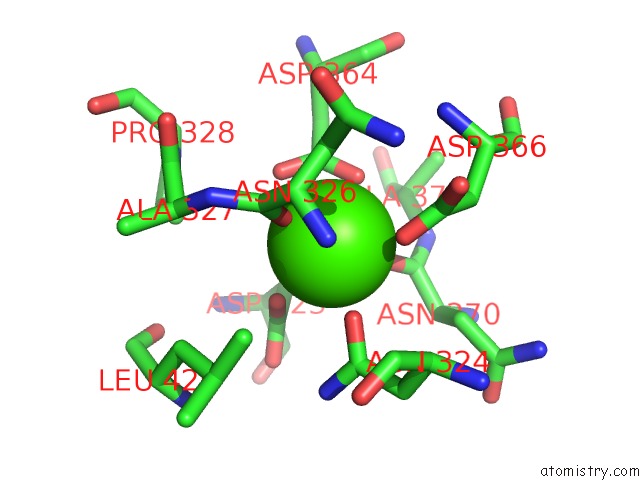
Calcium binding site 4 out of 9 in 6mga
Go back to
Calcium binding site 4 out
of 9 in the Crystal Structure of Human Protocadherin-1 EC1-4 with Glycosylation
Mono view

Stereo pair view
Mono view
Stereo pair view
A full contact list of Calcium with other atoms in the Ca binding
site number 4 of Crystal Structure of Human Protocadherin-1 EC1-4 with Glycosylation within 5.0Å range:
|
Calcium binding site 5 out of 9 in 6mga
Go back to
Calcium binding site 5 out
of 9 in the Crystal Structure of Human Protocadherin-1 EC1-4 with Glycosylation
Mono view
Stereo pair view
Mono view
Stereo pair view
A full contact list of Calcium with other atoms in the Ca binding
site number 5 of Crystal Structure of Human Protocadherin-1 EC1-4 with Glycosylation within 5.0Å range:
|
Calcium binding site 6 out of 9 in 6mga
Go back to
Calcium binding site 6 out
of 9 in the Crystal Structure of Human Protocadherin-1 EC1-4 with Glycosylation
Mono view
Stereo pair view
Mono view
Stereo pair view
A full contact list of Calcium with other atoms in the Ca binding
site number 6 of Crystal Structure of Human Protocadherin-1 EC1-4 with Glycosylation within 5.0Å range:
|
Calcium binding site 7 out of 9 in 6mga
Go back to
Calcium binding site 7 out
of 9 in the Crystal Structure of Human Protocadherin-1 EC1-4 with Glycosylation
Mono view
Stereo pair view
Mono view
Stereo pair view
A full contact list of Calcium with other atoms in the Ca binding
site number 7 of Crystal Structure of Human Protocadherin-1 EC1-4 with Glycosylation within 5.0Å range:
|
Calcium binding site 8 out of 9 in 6mga
Go back to
Calcium binding site 8 out
of 9 in the Crystal Structure of Human Protocadherin-1 EC1-4 with Glycosylation
Mono view
Stereo pair view
Mono view
Stereo pair view
A full contact list of Calcium with other atoms in the Ca binding
site number 8 of Crystal Structure of Human Protocadherin-1 EC1-4 with Glycosylation within 5.0Å range:
|
Calcium binding site 9 out of 9 in 6mga
Go back to
Calcium binding site 9 out
of 9 in the Crystal Structure of Human Protocadherin-1 EC1-4 with Glycosylation
Mono view
Stereo pair view
Mono view
Stereo pair view
A full contact list of Calcium with other atoms in the Ca binding
site number 9 of Crystal Structure of Human Protocadherin-1 EC1-4 with Glycosylation within 5.0Å range:
|
Reference:
D.Modak,
M.Sotomayor.
Identification of An Adhesive Interface For the Non-Clustered Delta 1 Protocadherin-1 Involved in Respiratory Diseases. Commun Biol V. 2 354 2019.
ISSN: ESSN 2399-3642
PubMed: 31583286
DOI: 10.1038/S42003-019-0586-0
Page generated: Wed Jul 9 16:06:30 2025
ISSN: ESSN 2399-3642
PubMed: 31583286
DOI: 10.1038/S42003-019-0586-0
Last articles
Cl in 5J2ZCl in 5J3B
Cl in 5J2H
Cl in 5J2K
Cl in 5J2J
Cl in 5J2G
Cl in 5J2F
Cl in 5J2E
Cl in 5J2D
Cl in 5J2C