Calcium »
PDB 6m4s-6mrl »
6mjp »
Calcium in PDB 6mjp: Lptb(E163Q)Fgc From Vibrio Cholerae
Protein crystallography data
The structure of Lptb(E163Q)Fgc From Vibrio Cholerae, PDB code: 6mjp
was solved by
T.W.Owens,
D.Kahne,
A.C.Kruse,
with X-Ray Crystallography technique. A brief refinement statistics is given in the table below:
| Resolution Low / High (Å) | 49.27 / 2.85 |
| Space group | C 1 2 1 |
| Cell size a, b, c (Å), α, β, γ (°) | 167.354, 80.729, 202.992, 90.00, 112.18, 90.00 |
| R / Rfree (%) | 24.2 / 29.2 |
Other elements in 6mjp:
The structure of Lptb(E163Q)Fgc From Vibrio Cholerae also contains other interesting chemical elements:
| Chlorine | (Cl) | 4 atoms |
Calcium Binding Sites:
The binding sites of Calcium atom in the Lptb(E163Q)Fgc From Vibrio Cholerae
(pdb code 6mjp). This binding sites where shown within
5.0 Angstroms radius around Calcium atom.
In total 2 binding sites of Calcium where determined in the Lptb(E163Q)Fgc From Vibrio Cholerae, PDB code: 6mjp:
Jump to Calcium binding site number: 1; 2;
In total 2 binding sites of Calcium where determined in the Lptb(E163Q)Fgc From Vibrio Cholerae, PDB code: 6mjp:
Jump to Calcium binding site number: 1; 2;
Calcium binding site 1 out of 2 in 6mjp
Go back to
Calcium binding site 1 out
of 2 in the Lptb(E163Q)Fgc From Vibrio Cholerae
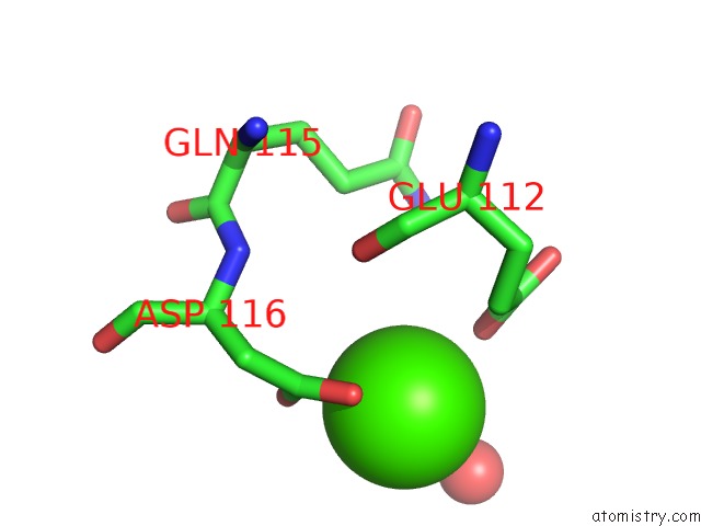
Mono view
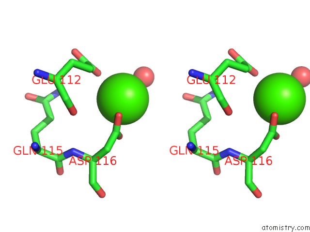
Stereo pair view
Mono view
Stereo pair view
A full contact list of Calcium with other atoms in the Ca binding
site number 1 of Lptb(E163Q)Fgc From Vibrio Cholerae within 5.0Å range:
|
Calcium binding site 2 out of 2 in 6mjp
Go back to
Calcium binding site 2 out
of 2 in the Lptb(E163Q)Fgc From Vibrio Cholerae
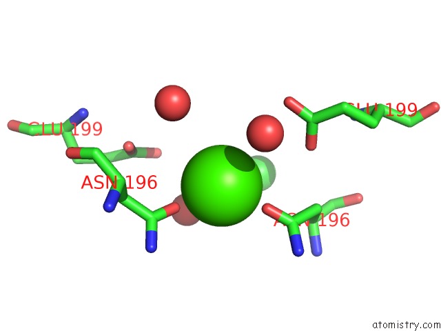
Mono view
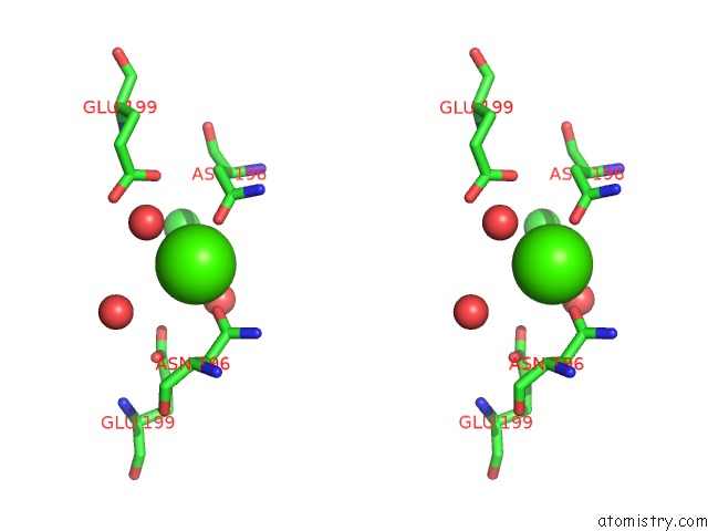
Stereo pair view
Mono view
Stereo pair view
A full contact list of Calcium with other atoms in the Ca binding
site number 2 of Lptb(E163Q)Fgc From Vibrio Cholerae within 5.0Å range:
|
Reference:
T.W.Owens,
R.J.Taylor,
K.S.Pahil,
B.R.Bertani,
N.Ruiz,
A.C.Kruse,
D.Kahne.
Structural Basis of Unidirectional Export of Lipopolysaccharide to the Cell Surface. Nature V. 567 550 2019.
ISSN: ESSN 1476-4687
PubMed: 30894747
DOI: 10.1038/S41586-019-1039-0
Page generated: Tue Jul 16 11:22:46 2024
ISSN: ESSN 1476-4687
PubMed: 30894747
DOI: 10.1038/S41586-019-1039-0
Last articles
Zn in 9MJ5Zn in 9HNW
Zn in 9G0L
Zn in 9FNE
Zn in 9DZN
Zn in 9E0I
Zn in 9D32
Zn in 9DAK
Zn in 8ZXC
Zn in 8ZUF