Calcium »
PDB 6mro-6n9d »
6mro »
Calcium in PDB 6mro: Crystal Structure of Methyl Transferase From Methanosarcina Acetivorans at 1.6 Angstroms Resolution, Northeast Structural Genomics Consortium (Nesg) Target MVR53.
Protein crystallography data
The structure of Crystal Structure of Methyl Transferase From Methanosarcina Acetivorans at 1.6 Angstroms Resolution, Northeast Structural Genomics Consortium (Nesg) Target MVR53., PDB code: 6mro
was solved by
S.Singh,
F.Forouhar,
C.Wang,
J.F.Hunt,
Northeast Structural Genomicsconsortium (Nesg),
with X-Ray Crystallography technique. A brief refinement statistics is given in the table below:
| Resolution Low / High (Å) | 40.99 / 1.60 |
| Space group | P 43 |
| Cell size a, b, c (Å), α, β, γ (°) | 40.990, 40.990, 104.850, 90.00, 90.00, 90.00 |
| R / Rfree (%) | 17.9 / 21 |
Other elements in 6mro:
The structure of Crystal Structure of Methyl Transferase From Methanosarcina Acetivorans at 1.6 Angstroms Resolution, Northeast Structural Genomics Consortium (Nesg) Target MVR53. also contains other interesting chemical elements:
| Chlorine | (Cl) | 8 atoms |
Calcium Binding Sites:
The binding sites of Calcium atom in the Crystal Structure of Methyl Transferase From Methanosarcina Acetivorans at 1.6 Angstroms Resolution, Northeast Structural Genomics Consortium (Nesg) Target MVR53.
(pdb code 6mro). This binding sites where shown within
5.0 Angstroms radius around Calcium atom.
In total 2 binding sites of Calcium where determined in the Crystal Structure of Methyl Transferase From Methanosarcina Acetivorans at 1.6 Angstroms Resolution, Northeast Structural Genomics Consortium (Nesg) Target MVR53., PDB code: 6mro:
Jump to Calcium binding site number: 1; 2;
In total 2 binding sites of Calcium where determined in the Crystal Structure of Methyl Transferase From Methanosarcina Acetivorans at 1.6 Angstroms Resolution, Northeast Structural Genomics Consortium (Nesg) Target MVR53., PDB code: 6mro:
Jump to Calcium binding site number: 1; 2;
Calcium binding site 1 out of 2 in 6mro
Go back to
Calcium binding site 1 out
of 2 in the Crystal Structure of Methyl Transferase From Methanosarcina Acetivorans at 1.6 Angstroms Resolution, Northeast Structural Genomics Consortium (Nesg) Target MVR53.
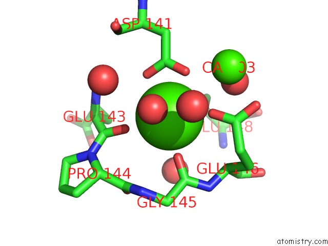
Mono view
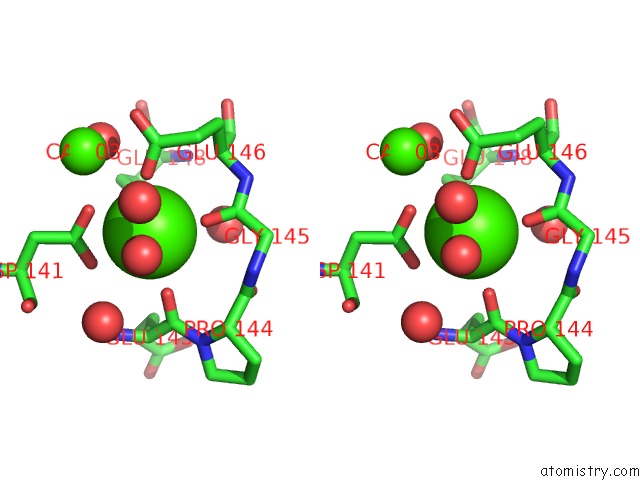
Stereo pair view
Mono view
Stereo pair view
A full contact list of Calcium with other atoms in the Ca binding
site number 1 of Crystal Structure of Methyl Transferase From Methanosarcina Acetivorans at 1.6 Angstroms Resolution, Northeast Structural Genomics Consortium (Nesg) Target MVR53. within 5.0Å range:
|
Calcium binding site 2 out of 2 in 6mro
Go back to
Calcium binding site 2 out
of 2 in the Crystal Structure of Methyl Transferase From Methanosarcina Acetivorans at 1.6 Angstroms Resolution, Northeast Structural Genomics Consortium (Nesg) Target MVR53.
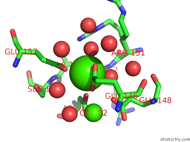
Mono view
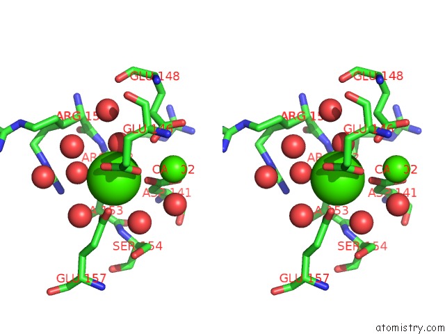
Stereo pair view
Mono view
Stereo pair view
A full contact list of Calcium with other atoms in the Ca binding
site number 2 of Crystal Structure of Methyl Transferase From Methanosarcina Acetivorans at 1.6 Angstroms Resolution, Northeast Structural Genomics Consortium (Nesg) Target MVR53. within 5.0Å range:
|
Reference:
S.Singh,
F.Forouhar,
C.Wang,
S.M.Vorobiev,
M.J.Neky,
J.F.Hunt.
Crystal Structure of A Methyl Transferase From Methanosarcina Acetivorans at 1.6 Angstroms Resolution. To Be Published.
Page generated: Wed Jul 9 16:10:06 2025
Last articles
Fe in 2YXOFe in 2YRS
Fe in 2YXC
Fe in 2YNM
Fe in 2YVJ
Fe in 2YP1
Fe in 2YU2
Fe in 2YU1
Fe in 2YQB
Fe in 2YOO