Calcium »
PDB 6mrm-6n8b »
6ms2 »
Calcium in PDB 6ms2: Crystal Structure of the GH43 Blxynb Protein From Bacillus Licheniformis
Protein crystallography data
The structure of Crystal Structure of the GH43 Blxynb Protein From Bacillus Licheniformis, PDB code: 6ms2
was solved by
L.M.Zanphorlin,
M.A.B.Morais,
J.A.Diogo,
M.T.Murakami,
with X-Ray Crystallography technique. A brief refinement statistics is given in the table below:
| Resolution Low / High (Å) | 33.31 / 2.49 |
| Space group | C 1 2 1 |
| Cell size a, b, c (Å), α, β, γ (°) | 152.858, 41.902, 71.805, 90.00, 91.70, 90.00 |
| R / Rfree (%) | 19.7 / 24.2 |
Calcium Binding Sites:
The binding sites of Calcium atom in the Crystal Structure of the GH43 Blxynb Protein From Bacillus Licheniformis
(pdb code 6ms2). This binding sites where shown within
5.0 Angstroms radius around Calcium atom.
In total only one binding site of Calcium was determined in the Crystal Structure of the GH43 Blxynb Protein From Bacillus Licheniformis, PDB code: 6ms2:
In total only one binding site of Calcium was determined in the Crystal Structure of the GH43 Blxynb Protein From Bacillus Licheniformis, PDB code: 6ms2:
Calcium binding site 1 out of 1 in 6ms2
Go back to
Calcium binding site 1 out
of 1 in the Crystal Structure of the GH43 Blxynb Protein From Bacillus Licheniformis
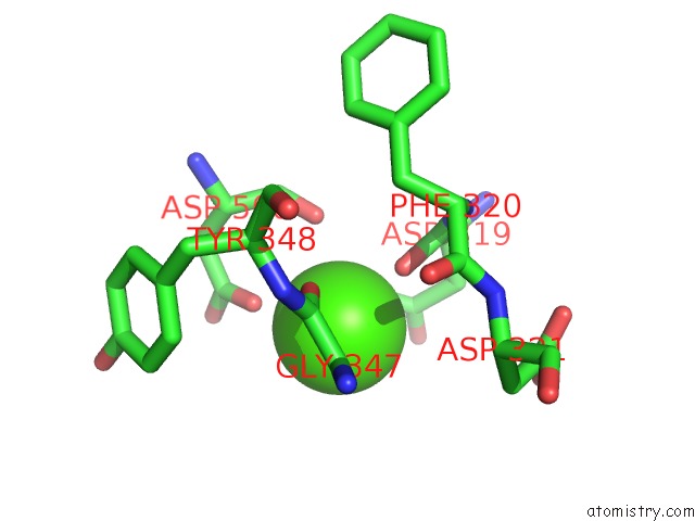
Mono view
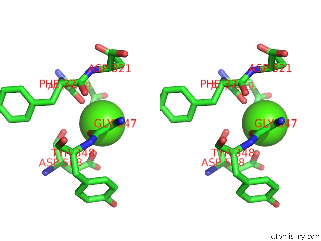
Stereo pair view
Mono view
Stereo pair view
A full contact list of Calcium with other atoms in the Ca binding
site number 1 of Crystal Structure of the GH43 Blxynb Protein From Bacillus Licheniformis within 5.0Å range:
|
Reference:
L.M.Zanphorlin,
M.A.B.De Morais,
J.A.Diogo,
M.N.Domingues,
F.H.M.De Souza,
R.Ruller,
M.T.Murakami.
Structure-Guided Design Combined with Evolutionary Diversity Led to the Discovery of the Xylose-Releasing Exo-Xylanase Activity in the Glycoside Hydrolase Family 43. Biotechnol. Bioeng. V. 116 734 2019.
ISSN: ESSN 1097-0290
PubMed: 30556897
DOI: 10.1002/BIT.26899
Page generated: Tue Jul 16 11:26:52 2024
ISSN: ESSN 1097-0290
PubMed: 30556897
DOI: 10.1002/BIT.26899
Last articles
Zn in 9MJ5Zn in 9HNW
Zn in 9G0L
Zn in 9FNE
Zn in 9DZN
Zn in 9E0I
Zn in 9D32
Zn in 9DAK
Zn in 8ZXC
Zn in 8ZUF