Calcium »
PDB 6mro-6n9d »
6mvh »
Calcium in PDB 6mvh: Crystal Structure of Fmn-Binding Beta-Glucuronidase From Roseburia Hominis
Enzymatic activity of Crystal Structure of Fmn-Binding Beta-Glucuronidase From Roseburia Hominis
All present enzymatic activity of Crystal Structure of Fmn-Binding Beta-Glucuronidase From Roseburia Hominis:
3.2.1.23;
3.2.1.23;
Protein crystallography data
The structure of Crystal Structure of Fmn-Binding Beta-Glucuronidase From Roseburia Hominis, PDB code: 6mvh
was solved by
S.J.Pellock,
M.R.Redinbo,
with X-Ray Crystallography technique. A brief refinement statistics is given in the table below:
| Resolution Low / High (Å) | 29.27 / 2.40 |
| Space group | P 1 21 1 |
| Cell size a, b, c (Å), α, β, γ (°) | 94.417, 137.407, 108.833, 90.00, 91.88, 90.00 |
| R / Rfree (%) | 20.9 / 26.8 |
Calcium Binding Sites:
The binding sites of Calcium atom in the Crystal Structure of Fmn-Binding Beta-Glucuronidase From Roseburia Hominis
(pdb code 6mvh). This binding sites where shown within
5.0 Angstroms radius around Calcium atom.
In total 4 binding sites of Calcium where determined in the Crystal Structure of Fmn-Binding Beta-Glucuronidase From Roseburia Hominis, PDB code: 6mvh:
Jump to Calcium binding site number: 1; 2; 3; 4;
In total 4 binding sites of Calcium where determined in the Crystal Structure of Fmn-Binding Beta-Glucuronidase From Roseburia Hominis, PDB code: 6mvh:
Jump to Calcium binding site number: 1; 2; 3; 4;
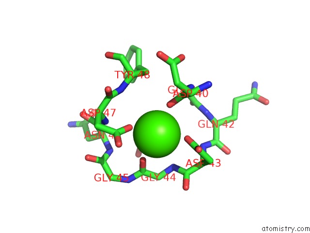
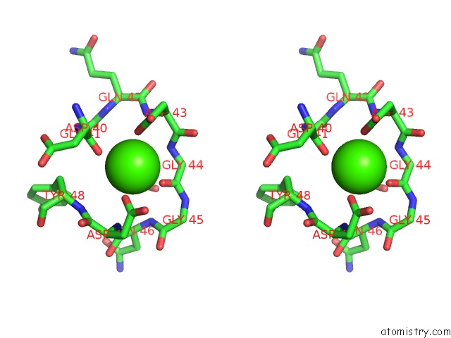
Calcium binding site 1 out of 4 in 6mvh
Go back to
Calcium binding site 1 out
of 4 in the Crystal Structure of Fmn-Binding Beta-Glucuronidase From Roseburia Hominis
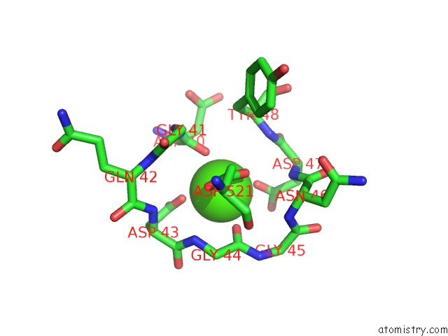
Mono view
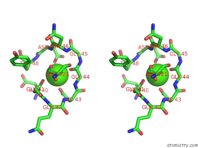
Stereo pair view
Mono view
Stereo pair view
A full contact list of Calcium with other atoms in the Ca binding
site number 1 of Crystal Structure of Fmn-Binding Beta-Glucuronidase From Roseburia Hominis within 5.0Å range:
|
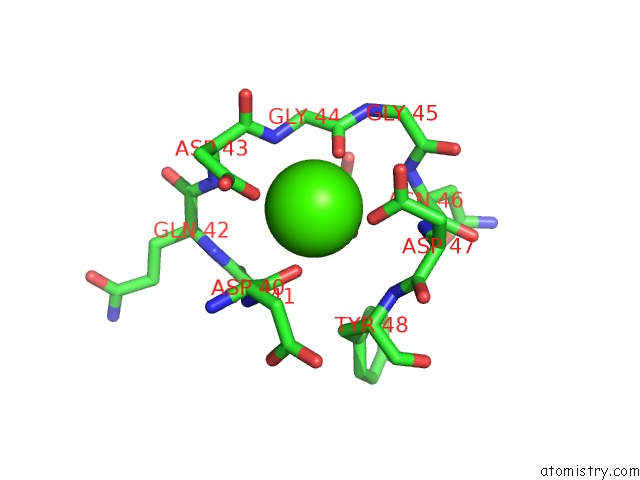
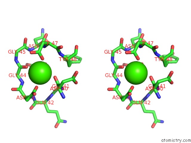
Calcium binding site 2 out of 4 in 6mvh
Go back to
Calcium binding site 2 out
of 4 in the Crystal Structure of Fmn-Binding Beta-Glucuronidase From Roseburia Hominis
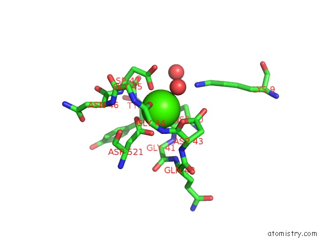
Mono view
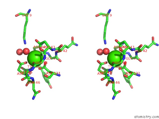
Stereo pair view
Mono view
Stereo pair view
A full contact list of Calcium with other atoms in the Ca binding
site number 2 of Crystal Structure of Fmn-Binding Beta-Glucuronidase From Roseburia Hominis within 5.0Å range:
|
Calcium binding site 3 out of 4 in 6mvh
Go back to
Calcium binding site 3 out
of 4 in the Crystal Structure of Fmn-Binding Beta-Glucuronidase From Roseburia Hominis
Mono view
Stereo pair view
Mono view
Stereo pair view
A full contact list of Calcium with other atoms in the Ca binding
site number 3 of Crystal Structure of Fmn-Binding Beta-Glucuronidase From Roseburia Hominis within 5.0Å range:
|
Calcium binding site 4 out of 4 in 6mvh
Go back to
Calcium binding site 4 out
of 4 in the Crystal Structure of Fmn-Binding Beta-Glucuronidase From Roseburia Hominis
Mono view
Stereo pair view
Mono view
Stereo pair view
A full contact list of Calcium with other atoms in the Ca binding
site number 4 of Crystal Structure of Fmn-Binding Beta-Glucuronidase From Roseburia Hominis within 5.0Å range:
|
Reference:
S.J.Pellock,
W.G.Walton,
S.M.Ervin,
D.Torres-Rivera,
B.C.Creekmore,
G.Bergan,
Z.D.Dunn,
B.Li,
A.Tripathy,
M.R.Redinbo.
Discovery and Characterization of Fmn-Binding Beta-Glucuronidases in the Human Gut Microbiome. J. Mol. Biol. V. 431 970 2019.
ISSN: ESSN 1089-8638
PubMed: 30658055
DOI: 10.1016/J.JMB.2019.01.013
Page generated: Wed Jul 9 16:11:43 2025
ISSN: ESSN 1089-8638
PubMed: 30658055
DOI: 10.1016/J.JMB.2019.01.013
Last articles
Ca in 7O3ACa in 7O1U
Ca in 7O1W
Ca in 7NSN
Ca in 7NZG
Ca in 7NZ6
Ca in 7NZ1
Ca in 7NYV
Ca in 7NYU
Ca in 7NYR