Calcium »
PDB 6mrm-6n8b »
6n1a »
Calcium in PDB 6n1a: Crystal Structure of An N-Acetylgalactosamine Deacetylase From F. Plautii
Protein crystallography data
The structure of Crystal Structure of An N-Acetylgalactosamine Deacetylase From F. Plautii, PDB code: 6n1a
was solved by
L.Sim,
P.Rahfeld,
S.G.Withers,
with X-Ray Crystallography technique. A brief refinement statistics is given in the table below:
| Resolution Low / High (Å) | 46.20 / 1.60 |
| Space group | P 21 21 21 |
| Cell size a, b, c (Å), α, β, γ (°) | 51.584, 69.186, 104.325, 90.00, 90.00, 90.00 |
| R / Rfree (%) | 13.9 / 16.7 |
Other elements in 6n1a:
The structure of Crystal Structure of An N-Acetylgalactosamine Deacetylase From F. Plautii also contains other interesting chemical elements:
| Manganese | (Mn) | 1 atom |
Calcium Binding Sites:
The binding sites of Calcium atom in the Crystal Structure of An N-Acetylgalactosamine Deacetylase From F. Plautii
(pdb code 6n1a). This binding sites where shown within
5.0 Angstroms radius around Calcium atom.
In total 2 binding sites of Calcium where determined in the Crystal Structure of An N-Acetylgalactosamine Deacetylase From F. Plautii, PDB code: 6n1a:
Jump to Calcium binding site number: 1; 2;
In total 2 binding sites of Calcium where determined in the Crystal Structure of An N-Acetylgalactosamine Deacetylase From F. Plautii, PDB code: 6n1a:
Jump to Calcium binding site number: 1; 2;
Calcium binding site 1 out of 2 in 6n1a
Go back to
Calcium binding site 1 out
of 2 in the Crystal Structure of An N-Acetylgalactosamine Deacetylase From F. Plautii
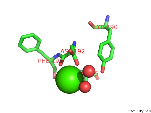
Mono view
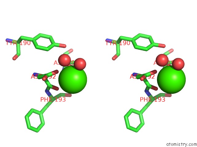
Stereo pair view
Mono view
Stereo pair view
A full contact list of Calcium with other atoms in the Ca binding
site number 1 of Crystal Structure of An N-Acetylgalactosamine Deacetylase From F. Plautii within 5.0Å range:
|
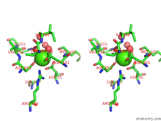
Calcium binding site 2 out of 2 in 6n1a
Go back to
Calcium binding site 2 out
of 2 in the Crystal Structure of An N-Acetylgalactosamine Deacetylase From F. Plautii
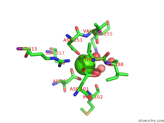
Mono view
Stereo pair view
Mono view
Stereo pair view
A full contact list of Calcium with other atoms in the Ca binding
site number 2 of Crystal Structure of An N-Acetylgalactosamine Deacetylase From F. Plautii within 5.0Å range:
|
Reference:
P.Rahfeld,
L.Sim,
H.Moon,
I.Constantinescu,
C.Morgan-Lang,
S.J.Hallam,
J.N.Kizhakkedathu,
S.G.Withers.
An Enzymatic Pathway in the Human Gut Microbiome That Converts A to Universal O Type Blood. Nat Microbiol V. 4 1475 2019.
ISSN: ESSN 2058-5276
PubMed: 31182795
DOI: 10.1038/S41564-019-0469-7
Page generated: Tue Jul 16 11:30:41 2024
ISSN: ESSN 2058-5276
PubMed: 31182795
DOI: 10.1038/S41564-019-0469-7
Last articles
Zn in 9MJ5Zn in 9HNW
Zn in 9G0L
Zn in 9FNE
Zn in 9DZN
Zn in 9E0I
Zn in 9D32
Zn in 9DAK
Zn in 8ZXC
Zn in 8ZUF