Calcium »
PDB 6mro-6n9d »
6n22 »
Calcium in PDB 6n22: Crystal Structure of Mouse Protocadherin-15 EC1-2 Bap
Protein crystallography data
The structure of Crystal Structure of Mouse Protocadherin-15 EC1-2 Bap, PDB code: 6n22
was solved by
Y.Narui,
M.Sotomayor,
with X-Ray Crystallography technique. A brief refinement statistics is given in the table below:
| Resolution Low / High (Å) | 49.86 / 2.40 |
| Space group | P 64 |
| Cell size a, b, c (Å), α, β, γ (°) | 99.616, 99.616, 58.560, 90.00, 90.00, 120.00 |
| R / Rfree (%) | 17 / 23.3 |
Calcium Binding Sites:
The binding sites of Calcium atom in the Crystal Structure of Mouse Protocadherin-15 EC1-2 Bap
(pdb code 6n22). This binding sites where shown within
5.0 Angstroms radius around Calcium atom.
In total 3 binding sites of Calcium where determined in the Crystal Structure of Mouse Protocadherin-15 EC1-2 Bap, PDB code: 6n22:
Jump to Calcium binding site number: 1; 2; 3;
In total 3 binding sites of Calcium where determined in the Crystal Structure of Mouse Protocadherin-15 EC1-2 Bap, PDB code: 6n22:
Jump to Calcium binding site number: 1; 2; 3;
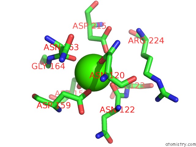
Calcium binding site 1 out of 3 in 6n22
Go back to
Calcium binding site 1 out
of 3 in the Crystal Structure of Mouse Protocadherin-15 EC1-2 Bap
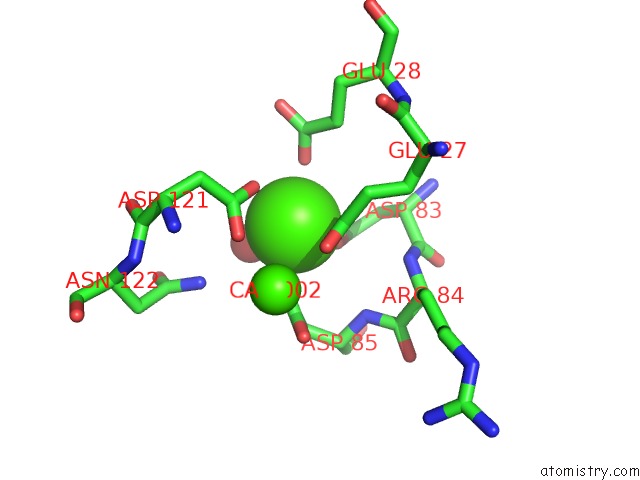
Mono view
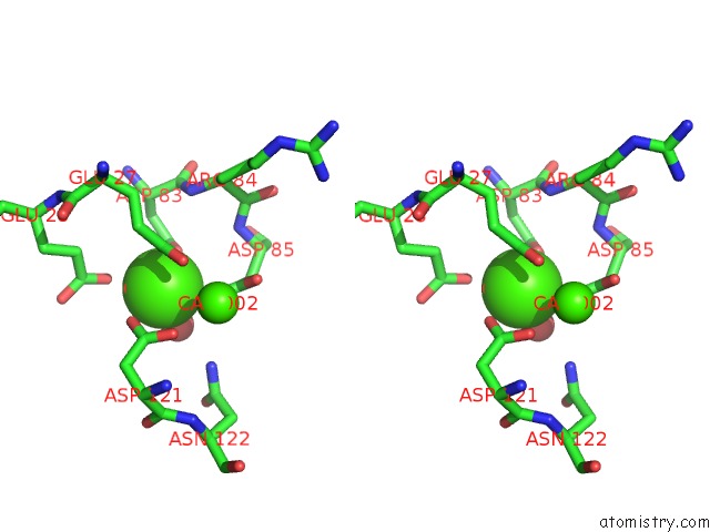
Stereo pair view
Mono view
Stereo pair view
A full contact list of Calcium with other atoms in the Ca binding
site number 1 of Crystal Structure of Mouse Protocadherin-15 EC1-2 Bap within 5.0Å range:
|
Calcium binding site 2 out of 3 in 6n22
Go back to
Calcium binding site 2 out
of 3 in the Crystal Structure of Mouse Protocadherin-15 EC1-2 Bap
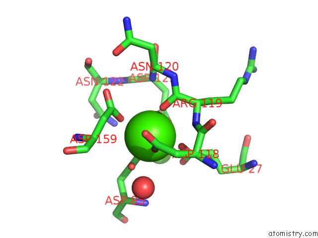
Mono view
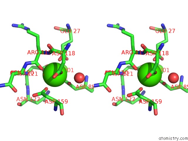
Stereo pair view
Mono view
Stereo pair view
A full contact list of Calcium with other atoms in the Ca binding
site number 2 of Crystal Structure of Mouse Protocadherin-15 EC1-2 Bap within 5.0Å range:
|
Calcium binding site 3 out of 3 in 6n22
Go back to
Calcium binding site 3 out
of 3 in the Crystal Structure of Mouse Protocadherin-15 EC1-2 Bap
Mono view

Stereo pair view
Mono view
Stereo pair view
A full contact list of Calcium with other atoms in the Ca binding
site number 3 of Crystal Structure of Mouse Protocadherin-15 EC1-2 Bap within 5.0Å range:
|
Reference:
Y.Narui,
M.Sotomayor.
Crystal Structure of Mouse Protocadherin-15 EC1-2 Bap To Be Published.
Page generated: Wed Jul 9 16:13:01 2025
Last articles
Ca in 7PC5Ca in 7PC4
Ca in 7PC3
Ca in 7PB7
Ca in 7PAG
Ca in 7PBY
Ca in 7PBA
Ca in 7PBB
Ca in 7P9T
Ca in 7PB5