Calcium »
PDB 6n9m-6o20 »
6n9m »
Calcium in PDB 6n9m: Crystal Structure of Adenosine Deaminase From Salmonella Typhimurium with Pentostatin (Deoxycoformycin)
Enzymatic activity of Crystal Structure of Adenosine Deaminase From Salmonella Typhimurium with Pentostatin (Deoxycoformycin)
All present enzymatic activity of Crystal Structure of Adenosine Deaminase From Salmonella Typhimurium with Pentostatin (Deoxycoformycin):
3.5.4.4;
3.5.4.4;
Protein crystallography data
The structure of Crystal Structure of Adenosine Deaminase From Salmonella Typhimurium with Pentostatin (Deoxycoformycin), PDB code: 6n9m
was solved by
N.Maltseva,
Y.Kim,
S.Grimshaw,
A.Joachimiak,
Center For Structuralgenomics Of Infectious Diseases (Csgid),
with X-Ray Crystallography technique. A brief refinement statistics is given in the table below:
| Resolution Low / High (Å) | 45.94 / 1.45 |
| Space group | P 21 21 21 |
| Cell size a, b, c (Å), α, β, γ (°) | 42.081, 73.232, 91.875, 90.00, 90.00, 90.00 |
| R / Rfree (%) | 15.5 / 16.8 |
Other elements in 6n9m:
The structure of Crystal Structure of Adenosine Deaminase From Salmonella Typhimurium with Pentostatin (Deoxycoformycin) also contains other interesting chemical elements:
| Zinc | (Zn) | 1 atom |
Calcium Binding Sites:
The binding sites of Calcium atom in the Crystal Structure of Adenosine Deaminase From Salmonella Typhimurium with Pentostatin (Deoxycoformycin)
(pdb code 6n9m). This binding sites where shown within
5.0 Angstroms radius around Calcium atom.
In total 2 binding sites of Calcium where determined in the Crystal Structure of Adenosine Deaminase From Salmonella Typhimurium with Pentostatin (Deoxycoformycin), PDB code: 6n9m:
Jump to Calcium binding site number: 1; 2;
In total 2 binding sites of Calcium where determined in the Crystal Structure of Adenosine Deaminase From Salmonella Typhimurium with Pentostatin (Deoxycoformycin), PDB code: 6n9m:
Jump to Calcium binding site number: 1; 2;
Calcium binding site 1 out of 2 in 6n9m
Go back to
Calcium binding site 1 out
of 2 in the Crystal Structure of Adenosine Deaminase From Salmonella Typhimurium with Pentostatin (Deoxycoformycin)
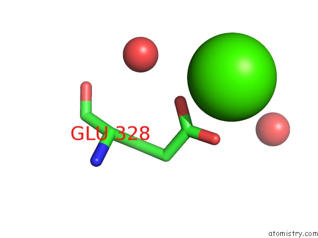
Mono view
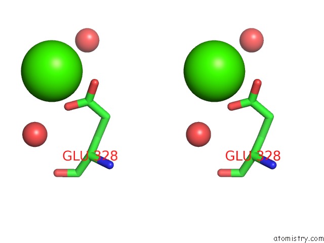
Stereo pair view
Mono view
Stereo pair view
A full contact list of Calcium with other atoms in the Ca binding
site number 1 of Crystal Structure of Adenosine Deaminase From Salmonella Typhimurium with Pentostatin (Deoxycoformycin) within 5.0Å range:
|
Calcium binding site 2 out of 2 in 6n9m
Go back to
Calcium binding site 2 out
of 2 in the Crystal Structure of Adenosine Deaminase From Salmonella Typhimurium with Pentostatin (Deoxycoformycin)
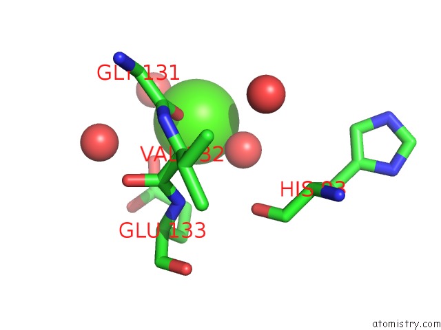
Mono view
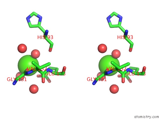
Stereo pair view
Mono view
Stereo pair view
A full contact list of Calcium with other atoms in the Ca binding
site number 2 of Crystal Structure of Adenosine Deaminase From Salmonella Typhimurium with Pentostatin (Deoxycoformycin) within 5.0Å range:
|
Reference:
N.Maltseva,
Y.Kim,
S.Grimshaw,
A.Joachimiak.
Crystal Structure of Adenosine Deaminase From Salmonella Typhimurium Complexed with Pentostatin (Deoxycoformycin) (Casp Target) To Be Published.
Page generated: Wed Jul 9 16:19:12 2025
Last articles
F in 7QM8F in 7QM7
F in 7QM5
F in 7QM4
F in 7QLT
F in 7QIT
F in 7QM3
F in 7QH1
F in 7QIS
F in 7QK4