Calcium »
PDB 6n9m-6o20 »
6nbt »
Calcium in PDB 6nbt: Crispr Complex Subunit CSM3 From Staphylococcus Epidermidis RP62A
Protein crystallography data
The structure of Crispr Complex Subunit CSM3 From Staphylococcus Epidermidis RP62A, PDB code: 6nbt
was solved by
B.W.Dorsey,
A.Mondragon,
with X-Ray Crystallography technique. A brief refinement statistics is given in the table below:
| Resolution Low / High (Å) | 25.00 / 2.40 |
| Space group | C 1 2 1 |
| Cell size a, b, c (Å), α, β, γ (°) | 104.612, 50.979, 83.018, 90.00, 97.05, 90.00 |
| R / Rfree (%) | 23 / 26.7 |
Other elements in 6nbt:
The structure of Crispr Complex Subunit CSM3 From Staphylococcus Epidermidis RP62A also contains other interesting chemical elements:
| Samarium | (Sm) | 13 atoms |
Calcium Binding Sites:
The binding sites of Calcium atom in the Crispr Complex Subunit CSM3 From Staphylococcus Epidermidis RP62A
(pdb code 6nbt). This binding sites where shown within
5.0 Angstroms radius around Calcium atom.
In total 3 binding sites of Calcium where determined in the Crispr Complex Subunit CSM3 From Staphylococcus Epidermidis RP62A, PDB code: 6nbt:
Jump to Calcium binding site number: 1; 2; 3;
In total 3 binding sites of Calcium where determined in the Crispr Complex Subunit CSM3 From Staphylococcus Epidermidis RP62A, PDB code: 6nbt:
Jump to Calcium binding site number: 1; 2; 3;
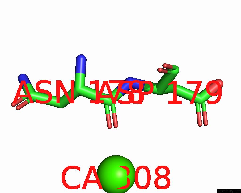
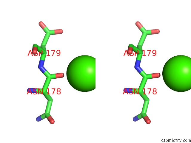
Calcium binding site 1 out of 3 in 6nbt
Go back to
Calcium binding site 1 out
of 3 in the Crispr Complex Subunit CSM3 From Staphylococcus Epidermidis RP62A
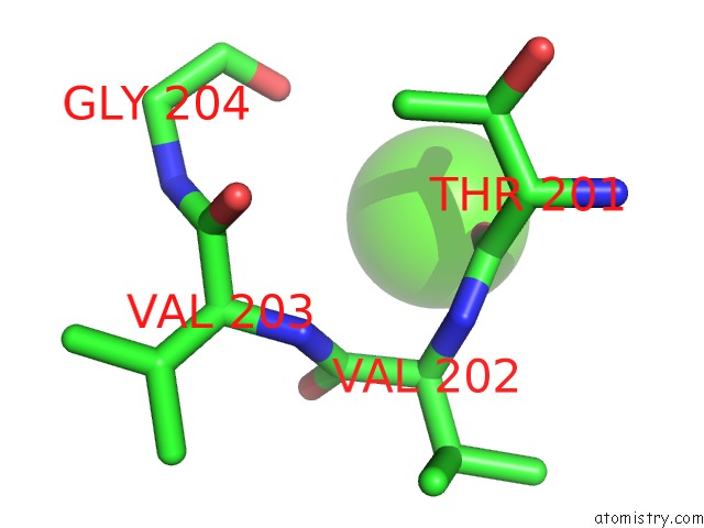
Mono view
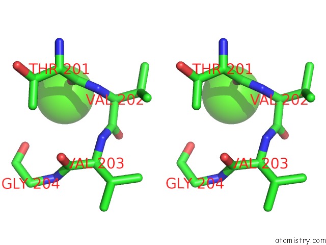
Stereo pair view
Mono view
Stereo pair view
A full contact list of Calcium with other atoms in the Ca binding
site number 1 of Crispr Complex Subunit CSM3 From Staphylococcus Epidermidis RP62A within 5.0Å range:
|
Calcium binding site 2 out of 3 in 6nbt
Go back to
Calcium binding site 2 out
of 3 in the Crispr Complex Subunit CSM3 From Staphylococcus Epidermidis RP62A
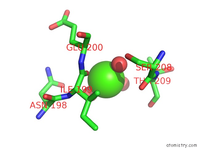
Mono view
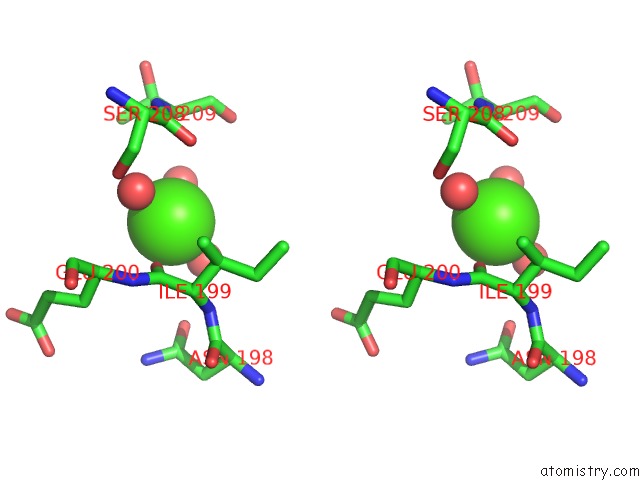
Stereo pair view
Mono view
Stereo pair view
A full contact list of Calcium with other atoms in the Ca binding
site number 2 of Crispr Complex Subunit CSM3 From Staphylococcus Epidermidis RP62A within 5.0Å range:
|
Calcium binding site 3 out of 3 in 6nbt
Go back to
Calcium binding site 3 out
of 3 in the Crispr Complex Subunit CSM3 From Staphylococcus Epidermidis RP62A
Mono view
Stereo pair view
Mono view
Stereo pair view
A full contact list of Calcium with other atoms in the Ca binding
site number 3 of Crispr Complex Subunit CSM3 From Staphylococcus Epidermidis RP62A within 5.0Å range:
|
Reference:
B.W.Dorsey,
L.Huang,
A.Mondragon.
Structural Organization of A Type III-A Crispr Effector Subcomplex Determined By X-Ray Crystallography and Cryo-Em. Nucleic Acids Res. V. 47 3765 2019.
ISSN: ESSN 1362-4962
PubMed: 30759237
DOI: 10.1093/NAR/GKZ079
Page generated: Wed Jul 9 16:19:31 2025
ISSN: ESSN 1362-4962
PubMed: 30759237
DOI: 10.1093/NAR/GKZ079
Last articles
Fe in 2YXOFe in 2YRS
Fe in 2YXC
Fe in 2YNM
Fe in 2YVJ
Fe in 2YP1
Fe in 2YU2
Fe in 2YU1
Fe in 2YQB
Fe in 2YOO