Calcium »
PDB 6n9m-6o20 »
6ncr »
Calcium in PDB 6ncr: Crystal Structure of Tryptophan-Trna Ligase From Chlamydia Trachomatis with Bound L-Tryptophan
Enzymatic activity of Crystal Structure of Tryptophan-Trna Ligase From Chlamydia Trachomatis with Bound L-Tryptophan
All present enzymatic activity of Crystal Structure of Tryptophan-Trna Ligase From Chlamydia Trachomatis with Bound L-Tryptophan:
6.1.1.2;
6.1.1.2;
Protein crystallography data
The structure of Crystal Structure of Tryptophan-Trna Ligase From Chlamydia Trachomatis with Bound L-Tryptophan, PDB code: 6ncr
was solved by
Seattle Structural Genomics Center For Infectious Disease,
Seattlestructural Genomics Center For Infectious Disease (Ssgcid),
with X-Ray Crystallography technique. A brief refinement statistics is given in the table below:
| Resolution Low / High (Å) | 47.03 / 1.75 |
| Space group | P 21 21 21 |
| Cell size a, b, c (Å), α, β, γ (°) | 50.890, 61.050, 246.160, 90.00, 90.00, 90.00 |
| R / Rfree (%) | 17.1 / 19.6 |
Calcium Binding Sites:
The binding sites of Calcium atom in the Crystal Structure of Tryptophan-Trna Ligase From Chlamydia Trachomatis with Bound L-Tryptophan
(pdb code 6ncr). This binding sites where shown within
5.0 Angstroms radius around Calcium atom.
In total only one binding site of Calcium was determined in the Crystal Structure of Tryptophan-Trna Ligase From Chlamydia Trachomatis with Bound L-Tryptophan, PDB code: 6ncr:
In total only one binding site of Calcium was determined in the Crystal Structure of Tryptophan-Trna Ligase From Chlamydia Trachomatis with Bound L-Tryptophan, PDB code: 6ncr:
Calcium binding site 1 out of 1 in 6ncr
Go back to
Calcium binding site 1 out
of 1 in the Crystal Structure of Tryptophan-Trna Ligase From Chlamydia Trachomatis with Bound L-Tryptophan
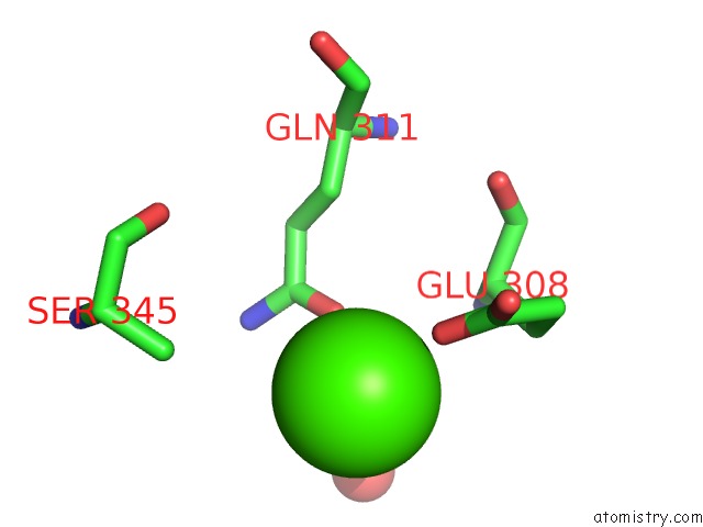
Mono view
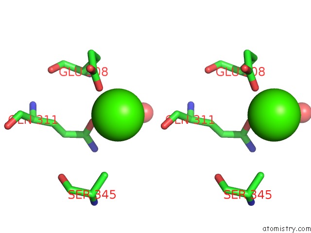
Stereo pair view
Mono view
Stereo pair view
A full contact list of Calcium with other atoms in the Ca binding
site number 1 of Crystal Structure of Tryptophan-Trna Ligase From Chlamydia Trachomatis with Bound L-Tryptophan within 5.0Å range:
|
Reference:
D.M.Dranow,
S.J.Mayclin,
D.D.Lorimer,
P.S.Horanyi,
T.E.Edwards.
Crystal Structure of Tryptophan-Trna Ligase From Chlamydia Trachomatis with Bound L-Tryptophan To Be Published.
Page generated: Wed Jul 9 16:19:52 2025
Last articles
F in 4FFWF in 4FM8
F in 4FM7
F in 4FLH
F in 4FIA
F in 4FKI
F in 4FK3
F in 4FJZ
F in 4FJY
F in 4FF6