Calcium in PDB 6oz7: Putative Oxidoreductase From Escherichia Coli Str. K-12
Protein crystallography data
The structure of Putative Oxidoreductase From Escherichia Coli Str. K-12, PDB code: 6oz7
was solved by
J.Osipiuk,
T.Skarina,
N.Mesa,
M.Endres,
A.Savchenko,
A.Joachimiak,
Centerfor Structural Genomics Of Infectious Diseases (Csgid),
with X-Ray Crystallography technique. A brief refinement statistics is given in the table below:
Resolution Low / High (Å)
|
48.14 /
1.36
|
Space group
|
P 1
|
Cell size a, b, c (Å), α, β, γ (°)
|
54.819,
72.210,
119.945,
82.59,
87.03,
67.82
|
R / Rfree (%)
|
12.3 /
16.9
|
Calcium Binding Sites:
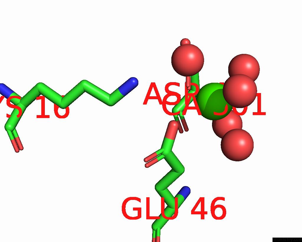
The binding sites of Calcium atom in the Putative Oxidoreductase From Escherichia Coli Str. K-12
(pdb code 6oz7). This binding sites where shown within
5.0 Angstroms radius around Calcium atom.
In total 8 binding sites of Calcium where determined in the
Putative Oxidoreductase From Escherichia Coli Str. K-12, PDB code: 6oz7:
Jump to Calcium binding site number:
1;
2;
3;
4;
5;
6;
7;
8;
Calcium binding site 1 out
of 8 in 6oz7
Go back to
Calcium Binding Sites List in 6oz7
Calcium binding site 1 out
of 8 in the Putative Oxidoreductase From Escherichia Coli Str. K-12
 Mono view
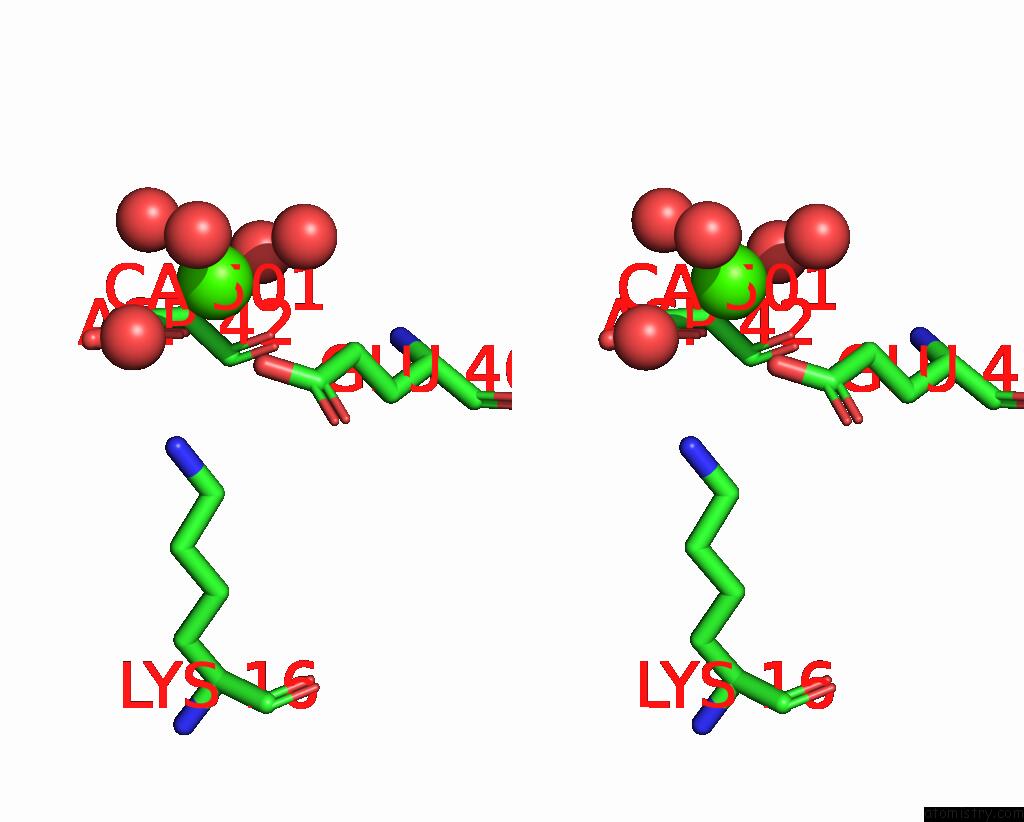
 Stereo pair view
|
A full contact list of Calcium with other atoms in the Ca binding
site number 1 of Putative Oxidoreductase From Escherichia Coli Str. K-12 within 5.0Å range:
probe
|
atom
|
residue
|
distance (Å)
|
B
|
Occ
|
A:Ca501
b:18.9
occ:0.80
|
OE2
|
A:GLU46
|
2.3
|
25.7
|
1.0
|
O
|
A:HOH760
|
2.3
|
32.8
|
1.0
|
OD1
|
A:ASP42
|
2.4
|
24.4
|
1.0
|
O
|
A:HOH776
|
2.4
|
20.8
|
1.0
|
O
|
A:HOH795
|
2.4
|
26.7
|
1.0
|
O
|
A:HOH672
|
2.5
|
25.2
|
1.0
|
O
|
A:HOH679
|
2.5
|
20.2
|
1.0
|
CG
|
A:ASP42
|
3.3
|
26.6
|
1.0
|
CD
|
A:GLU46
|
3.4
|
21.4
|
1.0
|
OD2
|
A:ASP42
|
3.7
|
38.2
|
1.0
|
CG
|
A:GLU46
|
3.8
|
21.2
|
1.0
|
NZ
|
A:LYS16
|
4.0
|
30.3
|
1.0
|
O
|
A:ASP42
|
4.3
|
17.6
|
1.0
|
CB
|
A:ASP42
|
4.5
|
22.5
|
1.0
|
OE1
|
A:GLU46
|
4.5
|
23.2
|
1.0
|
C
|
A:ASP42
|
4.7
|
17.8
|
1.0
|
CA
|
A:ASP42
|
4.8
|
19.8
|
1.0
|
|
Calcium binding site 2 out
of 8 in 6oz7
Go back to
Calcium Binding Sites List in 6oz7
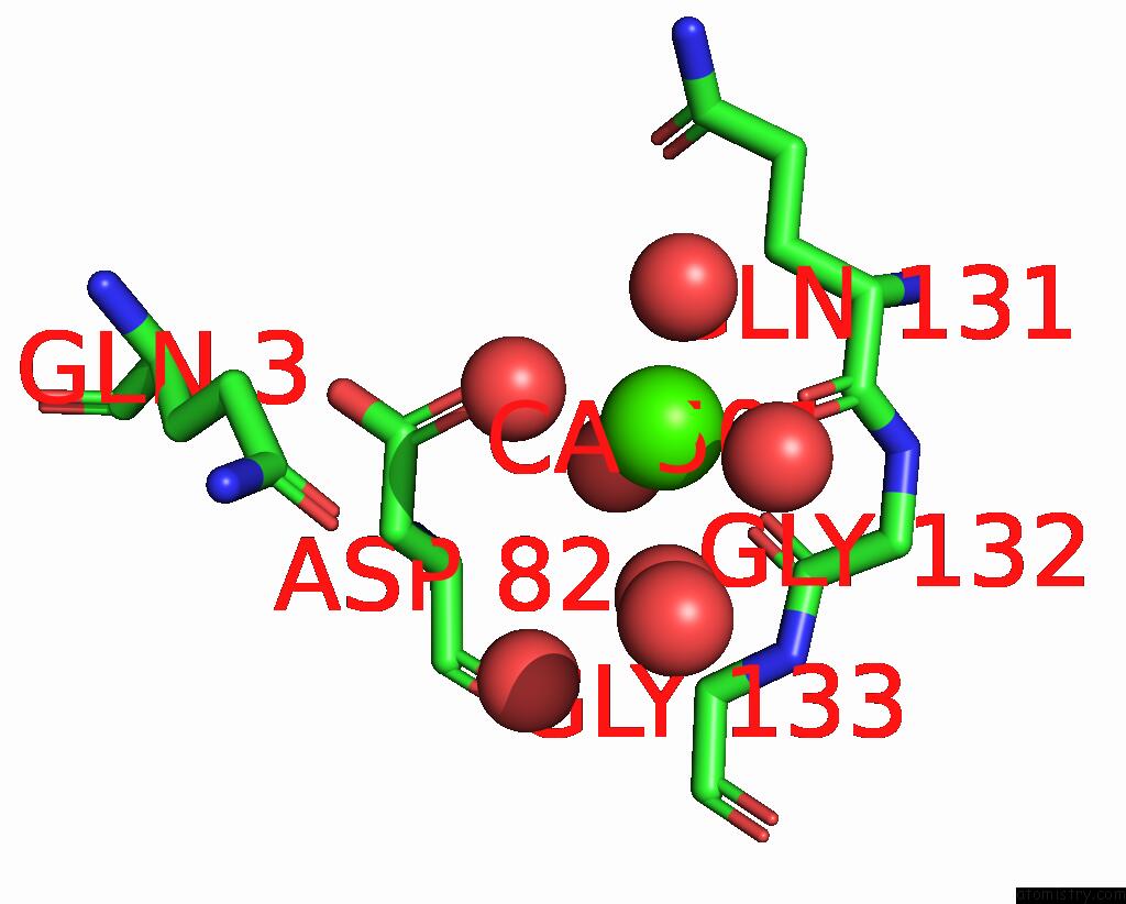
Calcium binding site 2 out
of 8 in the Putative Oxidoreductase From Escherichia Coli Str. K-12
 Mono view
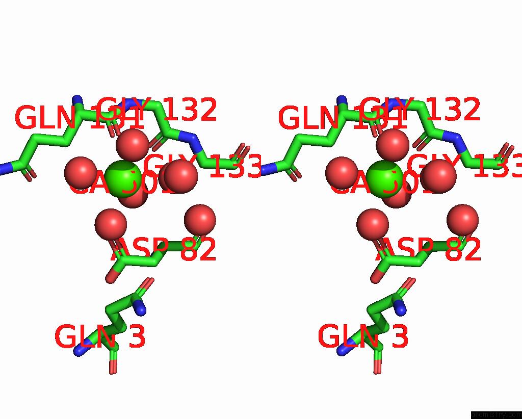
 Stereo pair view
|
A full contact list of Calcium with other atoms in the Ca binding
site number 2 of Putative Oxidoreductase From Escherichia Coli Str. K-12 within 5.0Å range:
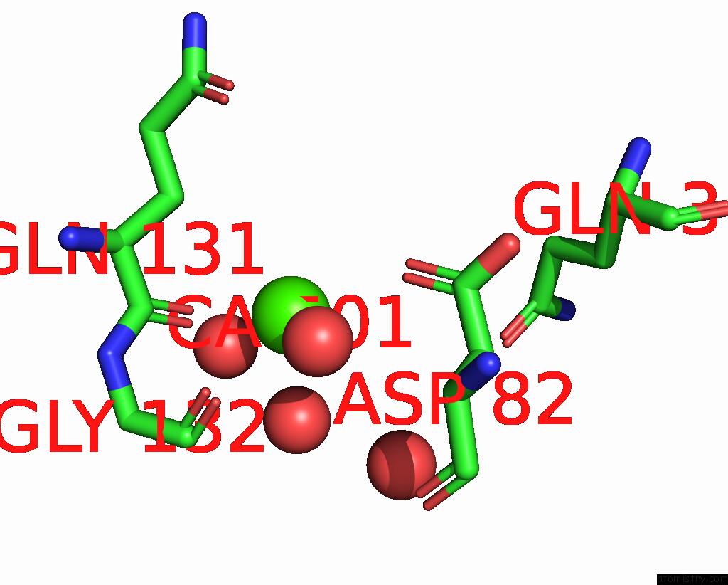
probe
|
atom
|
residue
|
distance (Å)
|
B
|
Occ
|
B:Ca501
b:28.2
occ:0.80
|
O
|
B:HOH774
|
2.2
|
42.7
|
1.0
|
O
|
B:HOH734
|
2.3
|
50.5
|
1.0
|
O
|
B:HOH747
|
2.3
|
39.0
|
1.0
|
O
|
B:HOH622
|
2.5
|
25.0
|
1.0
|
O
|
B:HOH785
|
2.5
|
29.4
|
1.0
|
O
|
B:GLN131
|
2.5
|
23.5
|
1.0
|
C
|
B:GLN131
|
3.7
|
22.3
|
1.0
|
O
|
B:HOH725
|
4.2
|
39.1
|
1.0
|
CB
|
B:GLN131
|
4.2
|
22.4
|
1.0
|
OD1
|
B:ASP82
|
4.3
|
18.9
|
1.0
|
O
|
B:HOH739
|
4.4
|
26.7
|
1.0
|
O
|
B:GLY132
|
4.4
|
16.5
|
1.0
|
C
|
B:GLY132
|
4.5
|
16.4
|
1.0
|
CA
|
B:GLN131
|
4.5
|
21.6
|
1.0
|
N
|
B:GLY132
|
4.6
|
18.6
|
1.0
|
CA
|
B:GLY132
|
4.7
|
18.4
|
1.0
|
N
|
B:GLY133
|
4.9
|
15.6
|
1.0
|
OE1
|
B:GLN3
|
5.0
|
38.4
|
1.0
|
|
Calcium binding site 3 out
of 8 in 6oz7
Go back to
Calcium Binding Sites List in 6oz7
Calcium binding site 3 out
of 8 in the Putative Oxidoreductase From Escherichia Coli Str. K-12
 Mono view
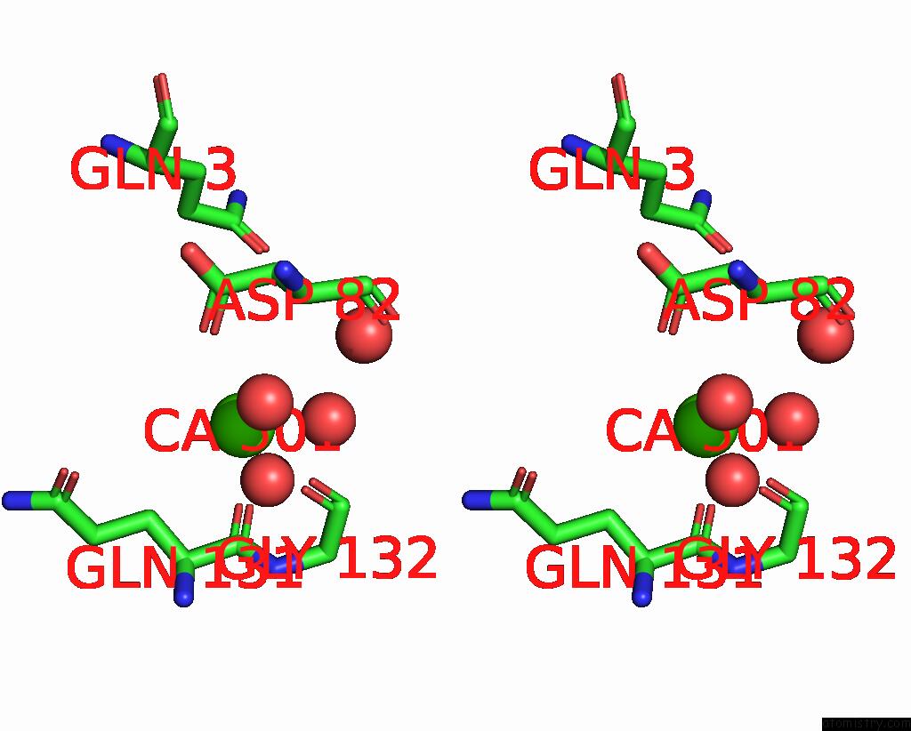
 Stereo pair view
|
A full contact list of Calcium with other atoms in the Ca binding
site number 3 of Putative Oxidoreductase From Escherichia Coli Str. K-12 within 5.0Å range:
probe
|
atom
|
residue
|
distance (Å)
|
B
|
Occ
|
C:Ca501
b:31.8
occ:0.70
|
O
|
C:HOH628
|
2.3
|
33.4
|
1.0
|
O
|
C:HOH760
|
2.4
|
36.8
|
1.0
|
O
|
C:GLN131
|
2.5
|
21.9
|
1.0
|
O
|
C:HOH617
|
2.5
|
28.2
|
1.0
|
C
|
C:GLN131
|
3.7
|
19.6
|
1.0
|
OD1
|
C:ASP82
|
4.3
|
19.9
|
1.0
|
CB
|
C:GLN131
|
4.3
|
20.7
|
1.0
|
O
|
C:HOH721
|
4.4
|
28.9
|
1.0
|
O
|
C:GLY132
|
4.5
|
18.2
|
1.0
|
CA
|
C:GLN131
|
4.5
|
19.4
|
1.0
|
C
|
C:GLY132
|
4.5
|
16.8
|
1.0
|
N
|
C:GLY132
|
4.7
|
17.7
|
1.0
|
CA
|
C:GLY132
|
4.7
|
17.3
|
1.0
|
OE1
|
C:GLN3
|
5.0
|
31.8
|
1.0
|
|
Calcium binding site 4 out
of 8 in 6oz7
Go back to
Calcium Binding Sites List in 6oz7
Calcium binding site 4 out
of 8 in the Putative Oxidoreductase From Escherichia Coli Str. K-12
 Mono view
 Stereo pair view
|
A full contact list of Calcium with other atoms in the Ca binding
site number 4 of Putative Oxidoreductase From Escherichia Coli Str. K-12 within 5.0Å range:
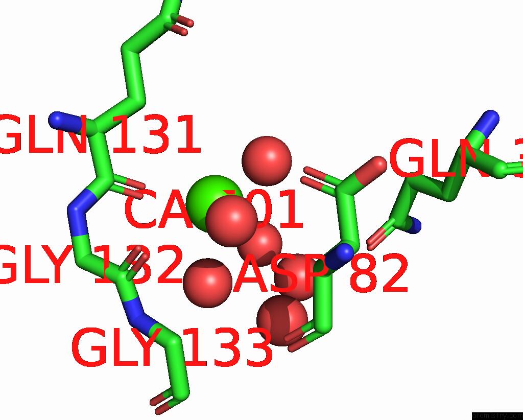
probe
|
atom
|
residue
|
distance (Å)
|
B
|
Occ
|
D:Ca501
b:17.9
occ:0.85
|
O
|
G:LYS128
|
2.3
|
20.4
|
1.0
|
O
|
G:HOH696
|
2.3
|
35.3
|
1.0
|
O
|
G:HOH704
|
2.3
|
23.7
|
1.0
|
O
|
G:HOH750
|
2.4
|
25.5
|
1.0
|
OE2
|
D:GLU37
|
2.4
|
21.4
|
1.0
|
O
|
D:HOH763
|
2.4
|
29.5
|
1.0
|
OE1
|
D:GLU37
|
2.5
|
19.0
|
1.0
|
CD
|
D:GLU37
|
2.8
|
19.6
|
1.0
|
C
|
G:LYS128
|
3.5
|
19.5
|
1.0
|
CA
|
G:LYS128
|
4.1
|
19.8
|
1.0
|
CG
|
D:GLU37
|
4.3
|
20.3
|
1.0
|
NZ
|
D:LYS41
|
4.4
|
36.2
|
1.0
|
O
|
D:HOH793
|
4.4
|
46.6
|
1.0
|
C
|
G:GLN129
|
4.4
|
21.0
|
1.0
|
O
|
G:GLN129
|
4.4
|
23.1
|
1.0
|
N
|
G:GLN129
|
4.5
|
19.2
|
1.0
|
O
|
D:HOH617
|
4.5
|
28.6
|
1.0
|
O
|
D:HOH692
|
4.6
|
23.6
|
1.0
|
O
|
D:HOH654
|
4.6
|
24.3
|
1.0
|
CZ2
|
D:TRP33
|
4.6
|
18.2
|
1.0
|
O
|
G:VAL127
|
4.7
|
20.1
|
1.0
|
CG
|
G:LYS128
|
4.8
|
29.6
|
1.0
|
CA
|
G:GLN129
|
4.8
|
20.3
|
1.0
|
O
|
D:HOH794
|
4.8
|
34.9
|
1.0
|
N
|
G:GLY130
|
4.8
|
22.2
|
1.0
|
O
|
D:HOH798
|
4.8
|
42.4
|
1.0
|
O
|
G:HOH774
|
4.8
|
42.8
|
1.0
|
O
|
G:HOH753
|
4.9
|
43.4
|
1.0
|
|
Calcium binding site 5 out
of 8 in 6oz7
Go back to
Calcium Binding Sites List in 6oz7
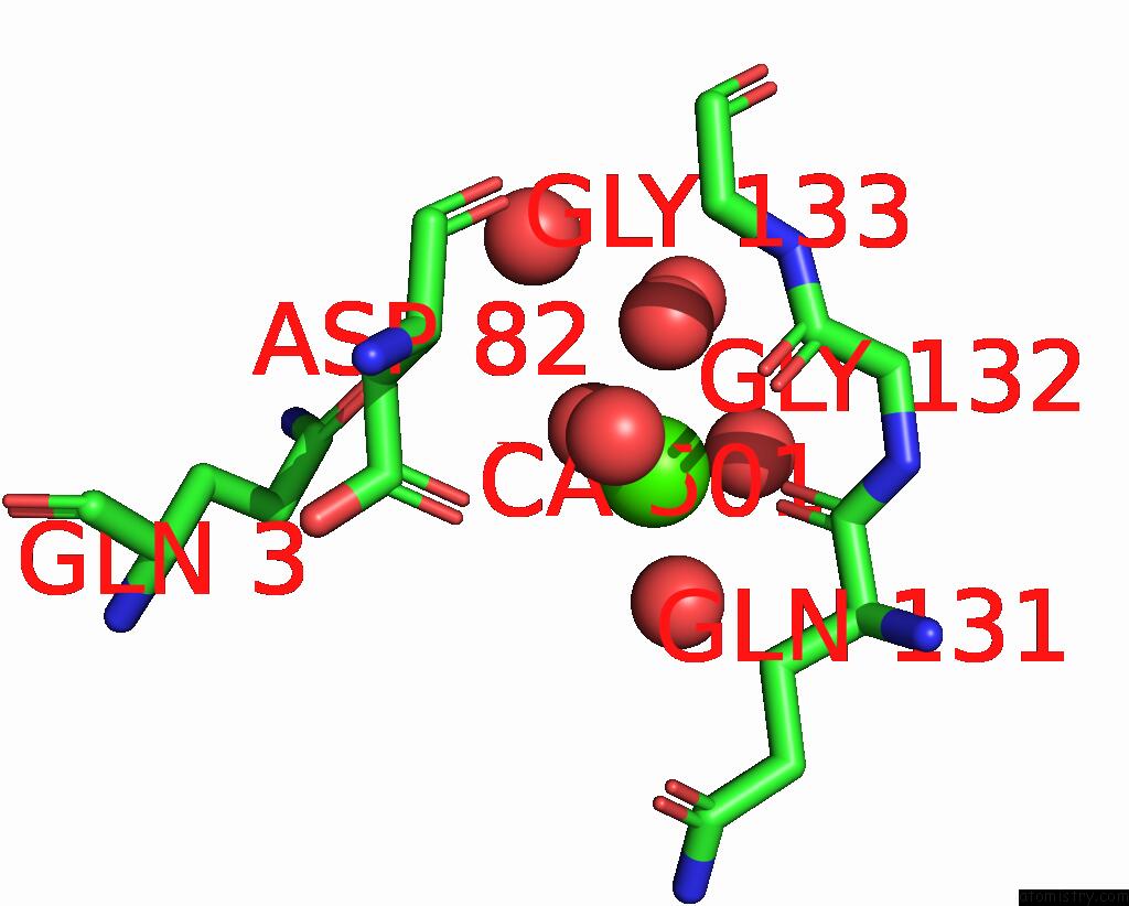
Calcium binding site 5 out
of 8 in the Putative Oxidoreductase From Escherichia Coli Str. K-12
 Mono view
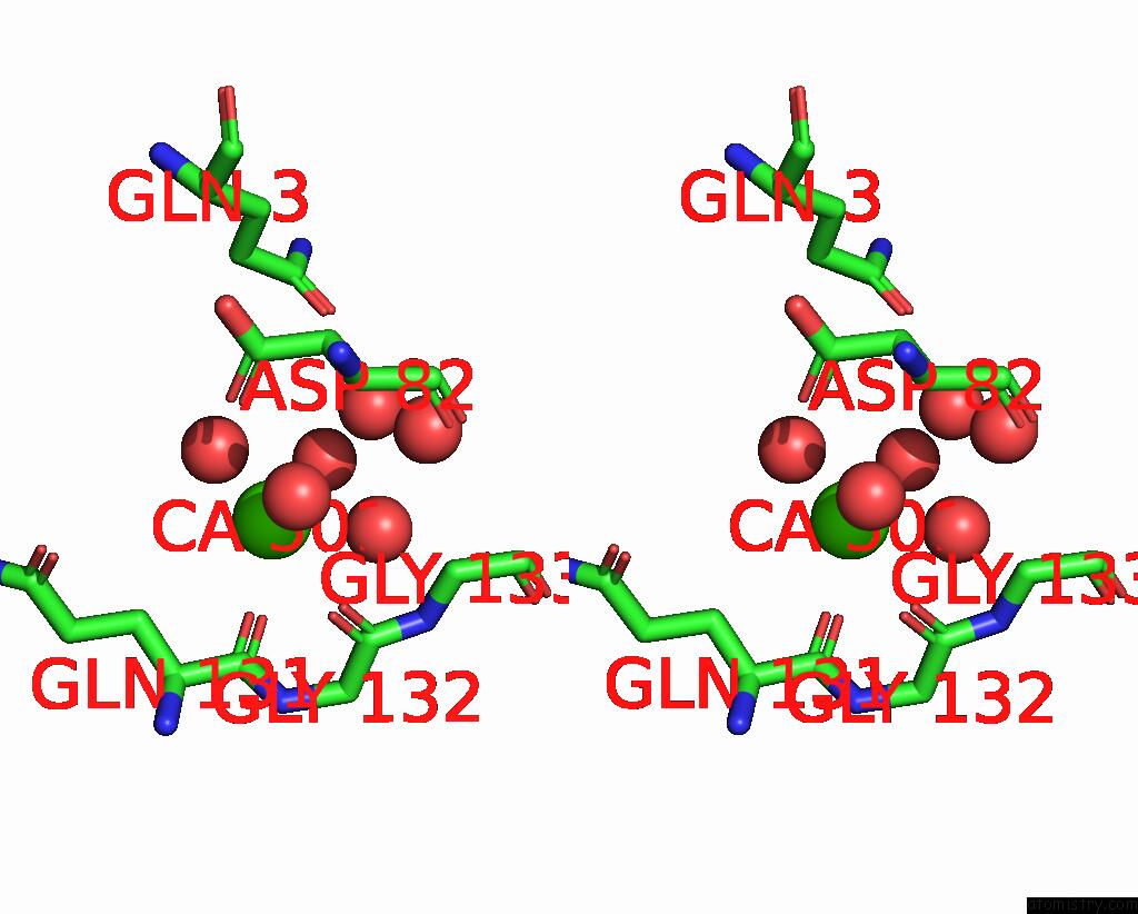
 Stereo pair view
|
A full contact list of Calcium with other atoms in the Ca binding
site number 5 of Putative Oxidoreductase From Escherichia Coli Str. K-12 within 5.0Å range:
probe
|
atom
|
residue
|
distance (Å)
|
B
|
Occ
|
E:Ca501
b:31.6
occ:0.70
|
O
|
E:HOH758
|
2.1
|
45.2
|
1.0
|
O
|
E:GLN131
|
2.5
|
24.6
|
1.0
|
O
|
E:HOH767
|
2.5
|
38.3
|
1.0
|
O
|
E:HOH624
|
2.5
|
27.2
|
1.0
|
C
|
E:GLN131
|
3.7
|
19.9
|
1.0
|
O
|
E:HOH604
|
3.8
|
22.7
|
1.0
|
O
|
E:HOH738
|
3.8
|
35.5
|
1.0
|
O
|
E:HOH682
|
4.3
|
37.0
|
1.0
|
CB
|
E:GLN131
|
4.3
|
24.7
|
1.0
|
OD1
|
E:ASP82
|
4.3
|
26.4
|
1.0
|
O
|
E:GLY132
|
4.4
|
19.8
|
1.0
|
C
|
E:GLY132
|
4.4
|
19.2
|
1.0
|
CA
|
E:GLN131
|
4.5
|
21.9
|
1.0
|
N
|
E:GLY132
|
4.6
|
19.9
|
1.0
|
CA
|
E:GLY132
|
4.6
|
18.4
|
1.0
|
N
|
E:GLY133
|
4.8
|
16.7
|
1.0
|
OE1
|
E:GLN3
|
5.0
|
39.2
|
1.0
|
|
Calcium binding site 6 out
of 8 in 6oz7
Go back to
Calcium Binding Sites List in 6oz7
Calcium binding site 6 out
of 8 in the Putative Oxidoreductase From Escherichia Coli Str. K-12
 Mono view
 Stereo pair view
|
A full contact list of Calcium with other atoms in the Ca binding
site number 6 of Putative Oxidoreductase From Escherichia Coli Str. K-12 within 5.0Å range:
probe
|
atom
|
residue
|
distance (Å)
|
B
|
Occ
|
F:Ca501
b:24.2
occ:0.70
|
O
|
F:HOH790
|
2.0
|
39.7
|
1.0
|
O
|
F:HOH753
|
2.3
|
41.8
|
1.0
|
O
|
F:HOH716
|
2.3
|
32.5
|
1.0
|
O
|
F:GLN131
|
2.4
|
22.6
|
1.0
|
O
|
F:HOH774
|
2.4
|
28.8
|
1.0
|
O
|
F:HOH651
|
2.5
|
25.9
|
1.0
|
C
|
F:GLN131
|
3.6
|
19.1
|
1.0
|
CB
|
F:GLN131
|
4.2
|
22.6
|
1.0
|
O
|
F:HOH768
|
4.2
|
38.0
|
1.0
|
O
|
F:HOH712
|
4.3
|
24.2
|
1.0
|
OD1
|
F:ASP82
|
4.4
|
20.8
|
1.0
|
O
|
F:GLY132
|
4.4
|
18.4
|
1.0
|
C
|
F:GLY132
|
4.4
|
16.5
|
1.0
|
CA
|
F:GLN131
|
4.5
|
21.1
|
1.0
|
N
|
F:GLY132
|
4.5
|
17.7
|
1.0
|
CA
|
F:GLY132
|
4.6
|
18.5
|
1.0
|
N
|
F:GLY133
|
4.8
|
15.8
|
1.0
|
OE1
|
F:GLN3
|
4.9
|
32.2
|
1.0
|
|
Calcium binding site 7 out
of 8 in 6oz7
Go back to
Calcium Binding Sites List in 6oz7
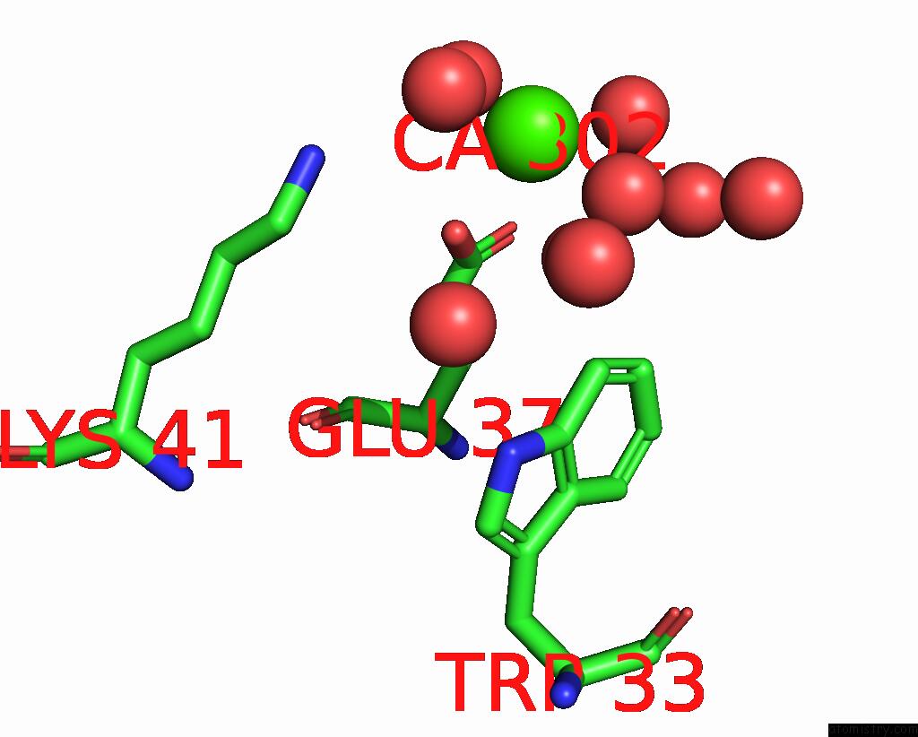
Calcium binding site 7 out
of 8 in the Putative Oxidoreductase From Escherichia Coli Str. K-12
 Mono view
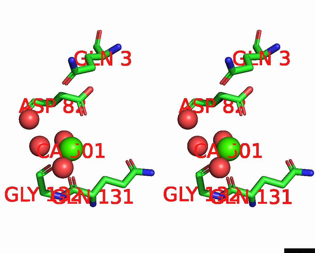
 Stereo pair view
|
A full contact list of Calcium with other atoms in the Ca binding
site number 7 of Putative Oxidoreductase From Escherichia Coli Str. K-12 within 5.0Å range:
probe
|
atom
|
residue
|
distance (Å)
|
B
|
Occ
|
G:Ca501
b:32.7
occ:0.70
|
O
|
G:HOH686
|
2.0
|
38.1
|
1.0
|
O
|
G:HOH758
|
2.4
|
42.4
|
1.0
|
O
|
G:GLN131
|
2.5
|
23.7
|
1.0
|
O
|
G:HOH641
|
2.5
|
25.2
|
1.0
|
C
|
G:GLN131
|
3.7
|
19.2
|
1.0
|
O
|
G:HOH726
|
4.2
|
33.4
|
1.0
|
CB
|
G:GLN131
|
4.3
|
24.0
|
1.0
|
OD1
|
G:ASP82
|
4.3
|
20.4
|
1.0
|
O
|
G:GLY132
|
4.5
|
18.9
|
1.0
|
C
|
G:GLY132
|
4.6
|
18.0
|
1.0
|
CA
|
G:GLN131
|
4.6
|
22.0
|
1.0
|
N
|
G:GLY132
|
4.7
|
19.0
|
1.0
|
CA
|
G:GLY132
|
4.8
|
20.8
|
1.0
|
OE1
|
G:GLN3
|
4.9
|
29.1
|
1.0
|
|
Calcium binding site 8 out
of 8 in 6oz7
Go back to
Calcium Binding Sites List in 6oz7
Calcium binding site 8 out
of 8 in the Putative Oxidoreductase From Escherichia Coli Str. K-12
 Mono view
 Stereo pair view
|
A full contact list of Calcium with other atoms in the Ca binding
site number 8 of Putative Oxidoreductase From Escherichia Coli Str. K-12 within 5.0Å range:
probe
|
atom
|
residue
|
distance (Å)
|
B
|
Occ
|
H:Ca302
b:17.9
occ:0.90
|
O
|
H:HOH600
|
2.3
|
22.9
|
1.0
|
O
|
H:HOH542
|
2.4
|
34.8
|
1.0
|
O
|
H:HOH588
|
2.4
|
24.0
|
1.0
|
OE2
|
H:GLU37
|
2.4
|
18.6
|
1.0
|
OE1
|
H:GLU37
|
2.5
|
18.6
|
1.0
|
O
|
H:HOH599
|
2.5
|
21.3
|
1.0
|
CD
|
H:GLU37
|
2.8
|
18.2
|
1.0
|
NZ
|
H:LYS41
|
4.2
|
30.2
|
1.0
|
CG
|
H:GLU37
|
4.3
|
18.2
|
1.0
|
O
|
H:HOH423
|
4.5
|
28.2
|
1.0
|
O
|
H:HOH481
|
4.6
|
22.1
|
1.0
|
O
|
H:HOH440
|
4.6
|
24.3
|
1.0
|
O
|
H:HOH624
|
4.6
|
31.7
|
1.0
|
CZ2
|
H:TRP33
|
4.7
|
16.9
|
1.0
|
O
|
H:HOH636
|
4.9
|
42.5
|
1.0
|
|
Reference:
J.Osipiuk,
T.Skarina,
N.Mesa,
M.Endres,
A.Savchenko,
A.Joachimiak,
Center For Structural Genomics Of Infectious Diseases(Csgid).
Putative Oxidoreductase From Escherichia Coli Str. K-12 To Be Published.
Page generated: Tue Jul 16 12:39:27 2024
|