Calcium »
PDB 6pu5-6q77 »
6pu5 »
Calcium in PDB 6pu5: Microed Structure of Proteinase K Recorded on Cetad
Enzymatic activity of Microed Structure of Proteinase K Recorded on Cetad
All present enzymatic activity of Microed Structure of Proteinase K Recorded on Cetad:
3.4.21.64;
3.4.21.64;
Calcium Binding Sites:
The binding sites of Calcium atom in the Microed Structure of Proteinase K Recorded on Cetad
(pdb code 6pu5). This binding sites where shown within
5.0 Angstroms radius around Calcium atom.
In total 2 binding sites of Calcium where determined in the Microed Structure of Proteinase K Recorded on Cetad, PDB code: 6pu5:
Jump to Calcium binding site number: 1; 2;
In total 2 binding sites of Calcium where determined in the Microed Structure of Proteinase K Recorded on Cetad, PDB code: 6pu5:
Jump to Calcium binding site number: 1; 2;
Calcium binding site 1 out of 2 in 6pu5
Go back to
Calcium binding site 1 out
of 2 in the Microed Structure of Proteinase K Recorded on Cetad
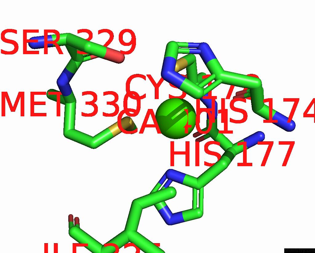
Mono view
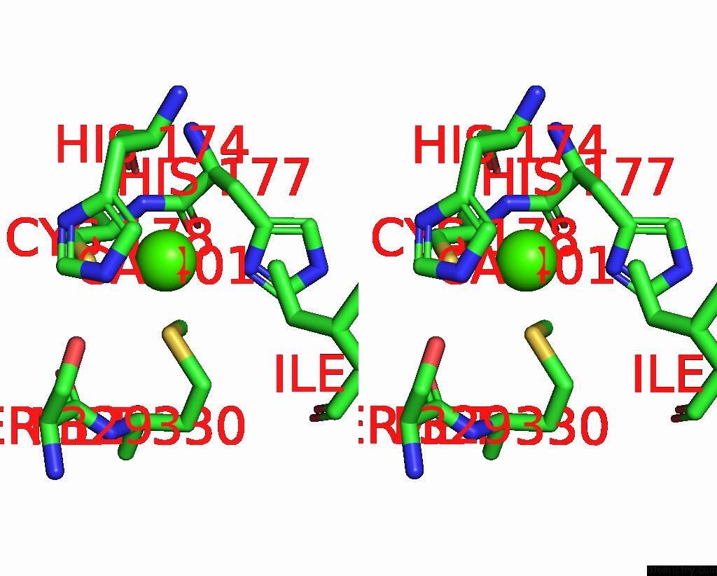
Stereo pair view
Mono view
Stereo pair view
A full contact list of Calcium with other atoms in the Ca binding
site number 1 of Microed Structure of Proteinase K Recorded on Cetad within 5.0Å range:
|
Calcium binding site 2 out of 2 in 6pu5
Go back to
Calcium binding site 2 out
of 2 in the Microed Structure of Proteinase K Recorded on Cetad
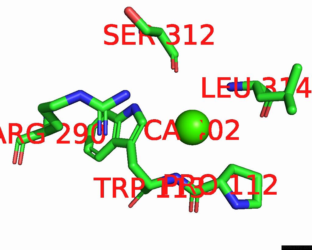
Mono view
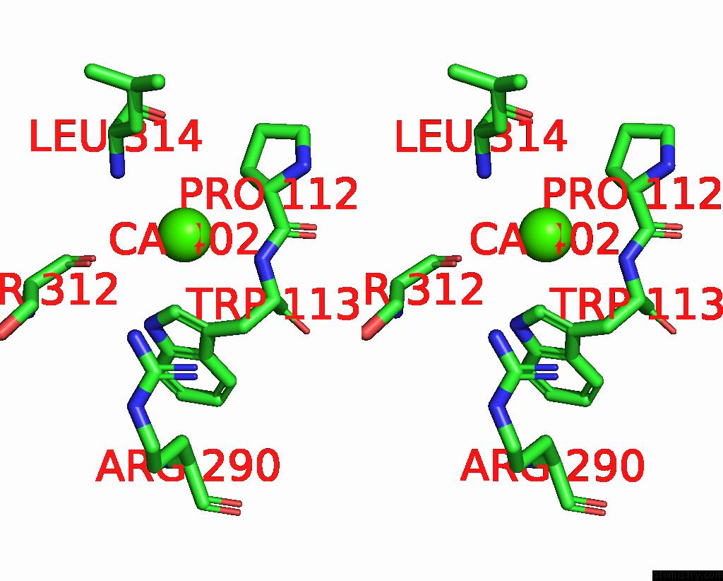
Stereo pair view
Mono view
Stereo pair view
A full contact list of Calcium with other atoms in the Ca binding
site number 2 of Microed Structure of Proteinase K Recorded on Cetad within 5.0Å range:
|
Reference:
J.Hattne,
M.W.Martynowycz,
P.A.Penczek,
T.Gonen.
Microed with the Falcon III Direct Electron Detector. Iucrj V. 6 921 2019.
ISSN: ESSN 2052-2525
PubMed: 31576224
DOI: 10.1107/S2052252519010583
Page generated: Tue Jul 16 13:05:44 2024
ISSN: ESSN 2052-2525
PubMed: 31576224
DOI: 10.1107/S2052252519010583
Last articles
Zn in 9J0NZn in 9J0O
Zn in 9J0P
Zn in 9FJX
Zn in 9EKB
Zn in 9C0F
Zn in 9CAH
Zn in 9CH0
Zn in 9CH3
Zn in 9CH1