Calcium »
PDB 6pu5-6q77 »
6q0c »
Calcium in PDB 6q0c: Muty Adenine Glycosylase Bound to Dna Containing A Transition State Analog (1N) Paired with Undamaged Dg
Enzymatic activity of Muty Adenine Glycosylase Bound to Dna Containing A Transition State Analog (1N) Paired with Undamaged Dg
All present enzymatic activity of Muty Adenine Glycosylase Bound to Dna Containing A Transition State Analog (1N) Paired with Undamaged Dg:
3.2.2.31;
3.2.2.31;
Protein crystallography data
The structure of Muty Adenine Glycosylase Bound to Dna Containing A Transition State Analog (1N) Paired with Undamaged Dg, PDB code: 6q0c
was solved by
V.L.O'shea Murray,
L.P.Russelburg,
M.P.Horvath,
S.S.David,
with X-Ray Crystallography technique. A brief refinement statistics is given in the table below:
| Resolution Low / High (Å) | 41.30 / 2.00 |
| Space group | P 21 21 21 |
| Cell size a, b, c (Å), α, β, γ (°) | 37.860, 86.140, 141.170, 90.00, 90.00, 90.00 |
| R / Rfree (%) | 24.7 / 27.1 |
Other elements in 6q0c:
The structure of Muty Adenine Glycosylase Bound to Dna Containing A Transition State Analog (1N) Paired with Undamaged Dg also contains other interesting chemical elements:
| Iron | (Fe) | 4 atoms |
Calcium Binding Sites:
The binding sites of Calcium atom in the Muty Adenine Glycosylase Bound to Dna Containing A Transition State Analog (1N) Paired with Undamaged Dg
(pdb code 6q0c). This binding sites where shown within
5.0 Angstroms radius around Calcium atom.
In total only one binding site of Calcium was determined in the Muty Adenine Glycosylase Bound to Dna Containing A Transition State Analog (1N) Paired with Undamaged Dg, PDB code: 6q0c:
In total only one binding site of Calcium was determined in the Muty Adenine Glycosylase Bound to Dna Containing A Transition State Analog (1N) Paired with Undamaged Dg, PDB code: 6q0c:
Calcium binding site 1 out of 1 in 6q0c
Go back to
Calcium binding site 1 out
of 1 in the Muty Adenine Glycosylase Bound to Dna Containing A Transition State Analog (1N) Paired with Undamaged Dg
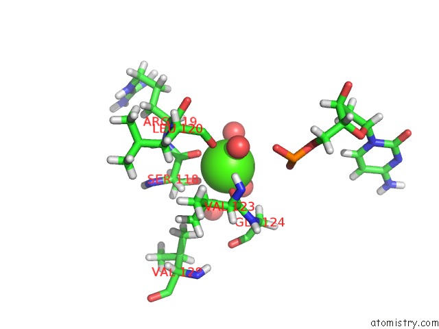
Mono view
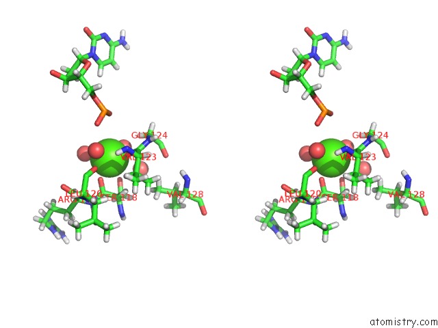
Stereo pair view
Mono view
Stereo pair view
A full contact list of Calcium with other atoms in the Ca binding
site number 1 of Muty Adenine Glycosylase Bound to Dna Containing A Transition State Analog (1N) Paired with Undamaged Dg within 5.0Å range:
|
Reference:
L.P.Russelburg,
V.L.O'shea Murray,
M.Demir,
K.R.Knutsen,
S.L.Sehgal,
S.Cao,
S.S.David,
M.P.Horvath.
Structural Basis For Finding Og Lesions and Avoiding Undamaged G By the Dna Glycosylase Muty To Be Published.
Page generated: Tue Jul 16 13:09:31 2024
Last articles
Zn in 9J0NZn in 9J0O
Zn in 9J0P
Zn in 9FJX
Zn in 9EKB
Zn in 9C0F
Zn in 9CAH
Zn in 9CH0
Zn in 9CH3
Zn in 9CH1