Calcium »
PDB 6pus-6q79 »
6q2y »
Calcium in PDB 6q2y: Crystal Structure of Ndm-1 Beta-Lactamase in Complex with Broad Spectrum Boronic Inhibitor CPD3
Enzymatic activity of Crystal Structure of Ndm-1 Beta-Lactamase in Complex with Broad Spectrum Boronic Inhibitor CPD3
All present enzymatic activity of Crystal Structure of Ndm-1 Beta-Lactamase in Complex with Broad Spectrum Boronic Inhibitor CPD3:
3.5.2.6;
3.5.2.6;
Protein crystallography data
The structure of Crystal Structure of Ndm-1 Beta-Lactamase in Complex with Broad Spectrum Boronic Inhibitor CPD3, PDB code: 6q2y
was solved by
L.Maso,
A.Quotadamo,
P.Bellio,
M.Montanari,
A.Venturelli,
G.Celenza,
M.P.Costi,
D.Tondi,
L.Cendron,
with X-Ray Crystallography technique. A brief refinement statistics is given in the table below:
| Resolution Low / High (Å) | 53.62 / 1.00 |
| Space group | P 21 21 21 |
| Cell size a, b, c (Å), α, β, γ (°) | 70.425, 73.950, 77.687, 90.00, 90.00, 90.00 |
| R / Rfree (%) | 16.6 / 17.6 |
Other elements in 6q2y:
The structure of Crystal Structure of Ndm-1 Beta-Lactamase in Complex with Broad Spectrum Boronic Inhibitor CPD3 also contains other interesting chemical elements:
| Zinc | (Zn) | 4 atoms |
Calcium Binding Sites:
The binding sites of Calcium atom in the Crystal Structure of Ndm-1 Beta-Lactamase in Complex with Broad Spectrum Boronic Inhibitor CPD3
(pdb code 6q2y). This binding sites where shown within
5.0 Angstroms radius around Calcium atom.
In total 4 binding sites of Calcium where determined in the Crystal Structure of Ndm-1 Beta-Lactamase in Complex with Broad Spectrum Boronic Inhibitor CPD3, PDB code: 6q2y:
Jump to Calcium binding site number: 1; 2; 3; 4;
In total 4 binding sites of Calcium where determined in the Crystal Structure of Ndm-1 Beta-Lactamase in Complex with Broad Spectrum Boronic Inhibitor CPD3, PDB code: 6q2y:
Jump to Calcium binding site number: 1; 2; 3; 4;
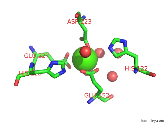
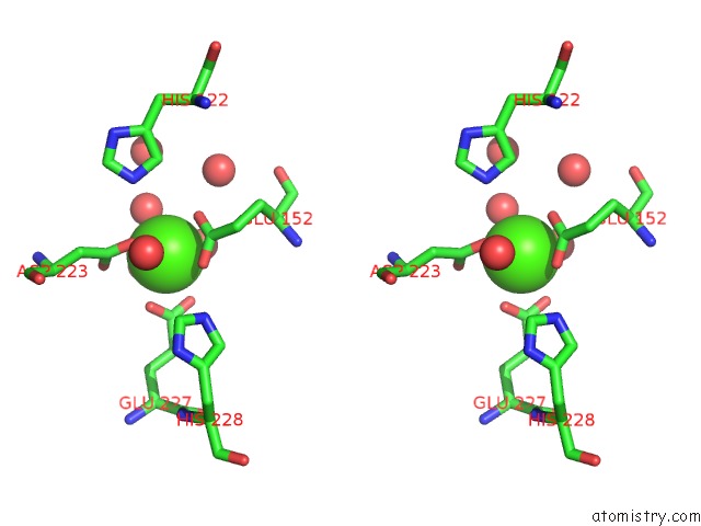
Calcium binding site 1 out of 4 in 6q2y
Go back to
Calcium binding site 1 out
of 4 in the Crystal Structure of Ndm-1 Beta-Lactamase in Complex with Broad Spectrum Boronic Inhibitor CPD3
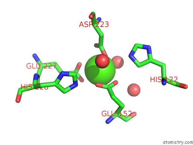
Mono view
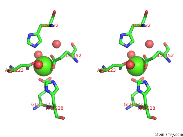
Stereo pair view
Mono view
Stereo pair view
A full contact list of Calcium with other atoms in the Ca binding
site number 1 of Crystal Structure of Ndm-1 Beta-Lactamase in Complex with Broad Spectrum Boronic Inhibitor CPD3 within 5.0Å range:
|
Calcium binding site 2 out of 4 in 6q2y
Go back to
Calcium binding site 2 out
of 4 in the Crystal Structure of Ndm-1 Beta-Lactamase in Complex with Broad Spectrum Boronic Inhibitor CPD3
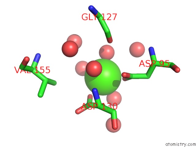
Mono view
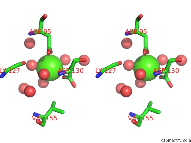
Stereo pair view
Mono view
Stereo pair view
A full contact list of Calcium with other atoms in the Ca binding
site number 2 of Crystal Structure of Ndm-1 Beta-Lactamase in Complex with Broad Spectrum Boronic Inhibitor CPD3 within 5.0Å range:
|
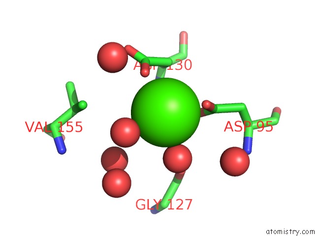
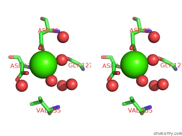
Calcium binding site 3 out of 4 in 6q2y
Go back to
Calcium binding site 3 out
of 4 in the Crystal Structure of Ndm-1 Beta-Lactamase in Complex with Broad Spectrum Boronic Inhibitor CPD3
Mono view
Stereo pair view
Mono view
Stereo pair view
A full contact list of Calcium with other atoms in the Ca binding
site number 3 of Crystal Structure of Ndm-1 Beta-Lactamase in Complex with Broad Spectrum Boronic Inhibitor CPD3 within 5.0Å range:
|
Calcium binding site 4 out of 4 in 6q2y
Go back to
Calcium binding site 4 out
of 4 in the Crystal Structure of Ndm-1 Beta-Lactamase in Complex with Broad Spectrum Boronic Inhibitor CPD3
Mono view
Stereo pair view
Mono view
Stereo pair view
A full contact list of Calcium with other atoms in the Ca binding
site number 4 of Crystal Structure of Ndm-1 Beta-Lactamase in Complex with Broad Spectrum Boronic Inhibitor CPD3 within 5.0Å range:
|
Reference:
L.Cendron,
A.Quotadamo,
L.Maso,
P.Bellio,
M.Montanari,
G.Celenza,
A.Venturelli,
M.P.Costi,
D.Tondi.
X-Ray Crystallography Deciphers the Activity of Broad-Spectrum Boronic Acid Beta-Lactamase Inhibitors. Acs Med.Chem.Lett. V. 10 650 2019.
ISSN: ISSN 1948-5875
PubMed: 30996812
DOI: 10.1021/ACSMEDCHEMLETT.8B00607
Page generated: Wed Jul 9 17:02:09 2025
ISSN: ISSN 1948-5875
PubMed: 30996812
DOI: 10.1021/ACSMEDCHEMLETT.8B00607
Last articles
Cl in 5JY1Cl in 5JYL
Cl in 5JXK
Cl in 5JXR
Cl in 5JXP
Cl in 5JXQ
Cl in 5JXJ
Cl in 5JX0
Cl in 5JXF
Cl in 5JX3