Calcium »
PDB 6qps-6r65 »
6qt6 »
Calcium in PDB 6qt6: Radiation Damage Study on A 16MER Dna Segment, Structure at 29.2 Mgy Dose
Protein crystallography data
The structure of Radiation Damage Study on A 16MER Dna Segment, Structure at 29.2 Mgy Dose, PDB code: 6qt6
was solved by
V.Bugris,
V.Harmat,
G.Ferenc,
S.Brockhauser,
I.Carmichael,
E.F.Garman,
with X-Ray Crystallography technique. A brief refinement statistics is given in the table below:
| Resolution Low / High (Å) | 31.43 / 1.80 |
| Space group | H 3 2 |
| Cell size a, b, c (Å), α, β, γ (°) | 36.965, 36.965, 162.571, 90.00, 90.00, 120.00 |
| R / Rfree (%) | 27 / 36.9 |
Calcium Binding Sites:
The binding sites of Calcium atom in the Radiation Damage Study on A 16MER Dna Segment, Structure at 29.2 Mgy Dose
(pdb code 6qt6). This binding sites where shown within
5.0 Angstroms radius around Calcium atom.
In total 6 binding sites of Calcium where determined in the Radiation Damage Study on A 16MER Dna Segment, Structure at 29.2 Mgy Dose, PDB code: 6qt6:
Jump to Calcium binding site number: 1; 2; 3; 4; 5; 6;
In total 6 binding sites of Calcium where determined in the Radiation Damage Study on A 16MER Dna Segment, Structure at 29.2 Mgy Dose, PDB code: 6qt6:
Jump to Calcium binding site number: 1; 2; 3; 4; 5; 6;
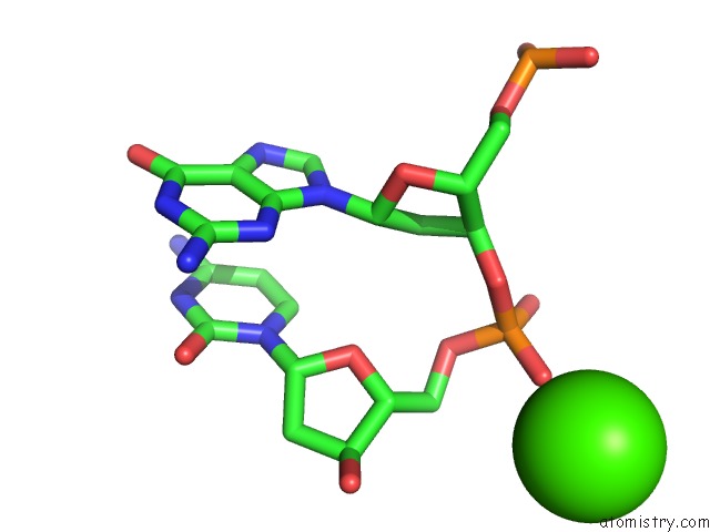
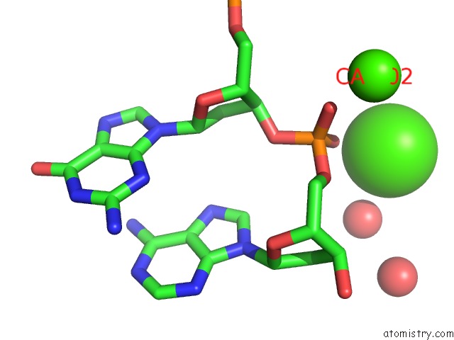
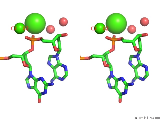
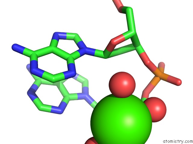
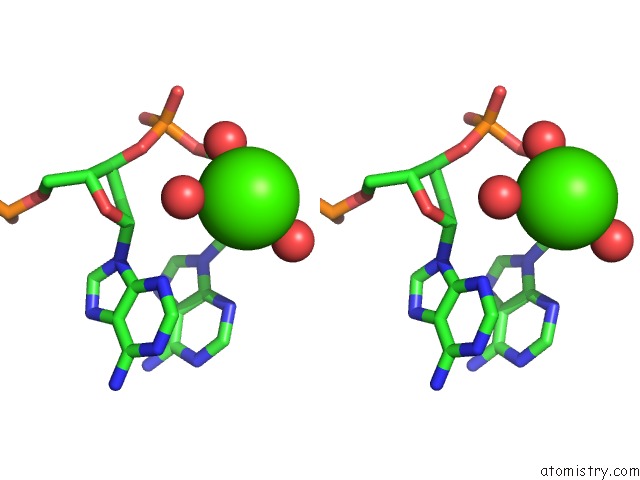
Calcium binding site 1 out of 6 in 6qt6
Go back to
Calcium binding site 1 out
of 6 in the Radiation Damage Study on A 16MER Dna Segment, Structure at 29.2 Mgy Dose
Mono view
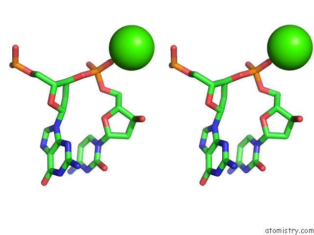
Stereo pair view
Mono view
Stereo pair view
A full contact list of Calcium with other atoms in the Ca binding
site number 1 of Radiation Damage Study on A 16MER Dna Segment, Structure at 29.2 Mgy Dose within 5.0Å range:
|
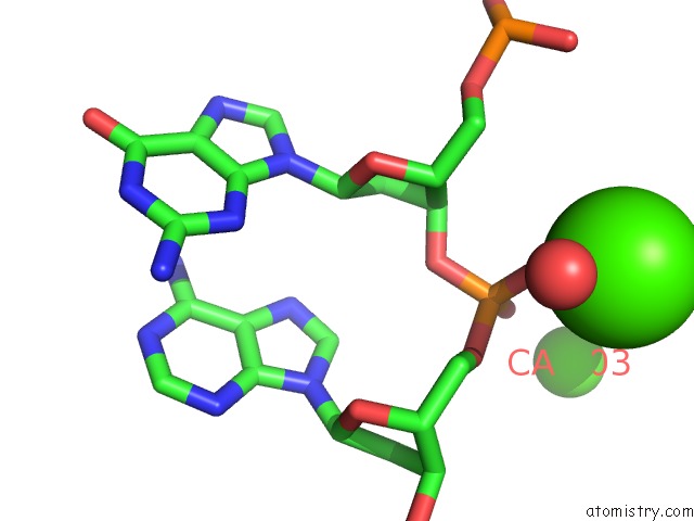
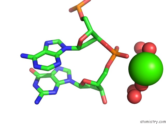
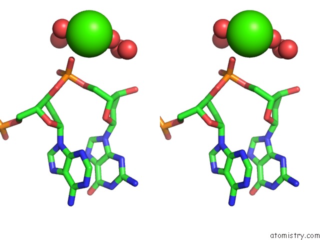
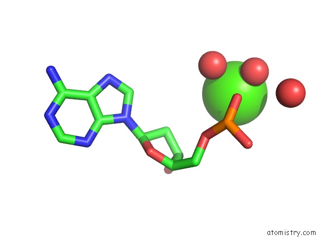
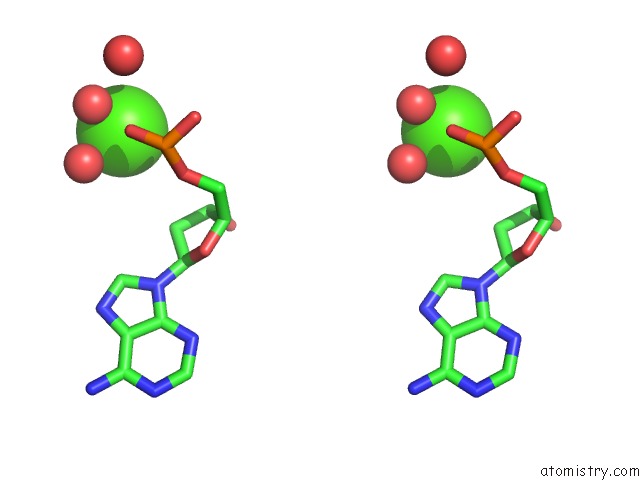
Calcium binding site 2 out of 6 in 6qt6
Go back to
Calcium binding site 2 out
of 6 in the Radiation Damage Study on A 16MER Dna Segment, Structure at 29.2 Mgy Dose
Mono view
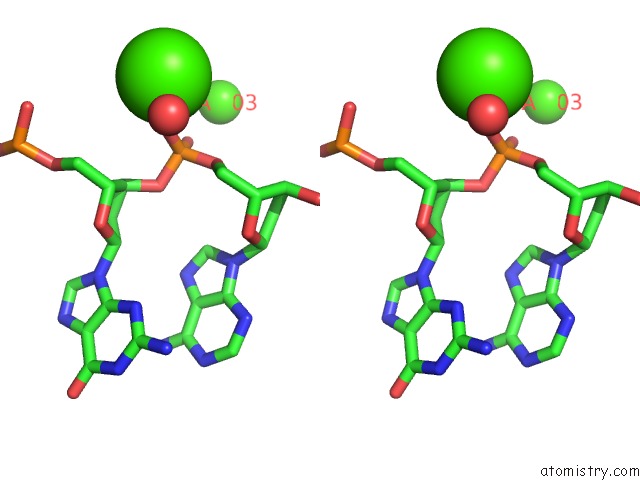
Stereo pair view
Mono view
Stereo pair view
A full contact list of Calcium with other atoms in the Ca binding
site number 2 of Radiation Damage Study on A 16MER Dna Segment, Structure at 29.2 Mgy Dose within 5.0Å range:
|
Calcium binding site 3 out of 6 in 6qt6
Go back to
Calcium binding site 3 out
of 6 in the Radiation Damage Study on A 16MER Dna Segment, Structure at 29.2 Mgy Dose
Mono view
Stereo pair view
Mono view
Stereo pair view
A full contact list of Calcium with other atoms in the Ca binding
site number 3 of Radiation Damage Study on A 16MER Dna Segment, Structure at 29.2 Mgy Dose within 5.0Å range:
|
Calcium binding site 4 out of 6 in 6qt6
Go back to
Calcium binding site 4 out
of 6 in the Radiation Damage Study on A 16MER Dna Segment, Structure at 29.2 Mgy Dose
Mono view
Stereo pair view
Mono view
Stereo pair view
A full contact list of Calcium with other atoms in the Ca binding
site number 4 of Radiation Damage Study on A 16MER Dna Segment, Structure at 29.2 Mgy Dose within 5.0Å range:
|
Calcium binding site 5 out of 6 in 6qt6
Go back to
Calcium binding site 5 out
of 6 in the Radiation Damage Study on A 16MER Dna Segment, Structure at 29.2 Mgy Dose
Mono view
Stereo pair view
Mono view
Stereo pair view
A full contact list of Calcium with other atoms in the Ca binding
site number 5 of Radiation Damage Study on A 16MER Dna Segment, Structure at 29.2 Mgy Dose within 5.0Å range:
|
Calcium binding site 6 out of 6 in 6qt6
Go back to
Calcium binding site 6 out
of 6 in the Radiation Damage Study on A 16MER Dna Segment, Structure at 29.2 Mgy Dose
Mono view
Stereo pair view
Mono view
Stereo pair view
A full contact list of Calcium with other atoms in the Ca binding
site number 6 of Radiation Damage Study on A 16MER Dna Segment, Structure at 29.2 Mgy Dose within 5.0Å range:
|
Reference:
V.Bugris,
V.Harmat,
G.Ferenc,
S.Brockhauser,
I.Carmichael,
E.F.Garman.
Radiation-Damage Investigation of A Dna 16-Mer. J.Synchrotron Radiat. V. 26 998 2019.
ISSN: ESSN 1600-5775
PubMed: 31274421
DOI: 10.1107/S160057751900763X
Page generated: Tue Jul 16 14:02:40 2024
ISSN: ESSN 1600-5775
PubMed: 31274421
DOI: 10.1107/S160057751900763X
Last articles
Zn in 9J0NZn in 9J0O
Zn in 9J0P
Zn in 9FJX
Zn in 9EKB
Zn in 9C0F
Zn in 9CAH
Zn in 9CH0
Zn in 9CH3
Zn in 9CH1