Calcium »
PDB 6qps-6r65 »
6r35 »
Calcium in PDB 6r35: Structure of the Lecb Lectin From Pseudomonas Aeruginosa Strain PAO1 in Complex with Lewis X Tetrasaccharide
Protein crystallography data
The structure of Structure of the Lecb Lectin From Pseudomonas Aeruginosa Strain PAO1 in Complex with Lewis X Tetrasaccharide, PDB code: 6r35
was solved by
M.Lepsik,
R.Sommer,
S.Kuhaudomlarp,
M.Lelimousin,
A.Varrot,
A.Titz,
A.Imberty,
with X-Ray Crystallography technique. A brief refinement statistics is given in the table below:
| Resolution Low / High (Å) | 47.87 / 1.80 |
| Space group | P 1 21 1 |
| Cell size a, b, c (Å), α, β, γ (°) | 52.603, 72.481, 62.061, 90.00, 114.62, 90.00 |
| R / Rfree (%) | 13.2 / 17.6 |
Calcium Binding Sites:
The binding sites of Calcium atom in the Structure of the Lecb Lectin From Pseudomonas Aeruginosa Strain PAO1 in Complex with Lewis X Tetrasaccharide
(pdb code 6r35). This binding sites where shown within
5.0 Angstroms radius around Calcium atom.
In total 8 binding sites of Calcium where determined in the Structure of the Lecb Lectin From Pseudomonas Aeruginosa Strain PAO1 in Complex with Lewis X Tetrasaccharide, PDB code: 6r35:
Jump to Calcium binding site number: 1; 2; 3; 4; 5; 6; 7; 8;
In total 8 binding sites of Calcium where determined in the Structure of the Lecb Lectin From Pseudomonas Aeruginosa Strain PAO1 in Complex with Lewis X Tetrasaccharide, PDB code: 6r35:
Jump to Calcium binding site number: 1; 2; 3; 4; 5; 6; 7; 8;
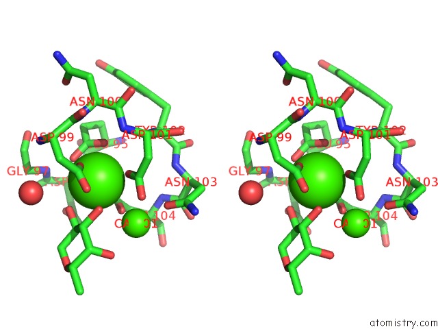
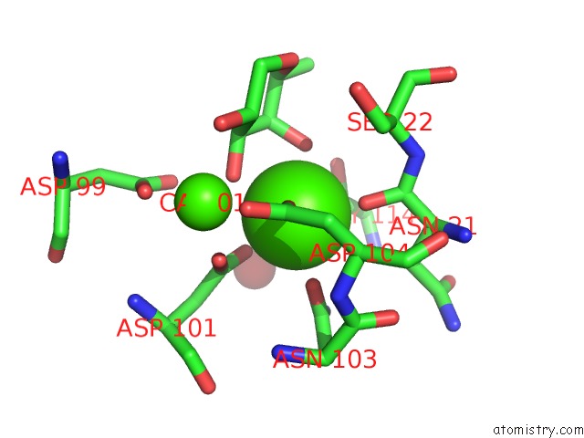
Calcium binding site 1 out of 8 in 6r35
Go back to
Calcium binding site 1 out
of 8 in the Structure of the Lecb Lectin From Pseudomonas Aeruginosa Strain PAO1 in Complex with Lewis X Tetrasaccharide
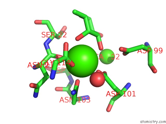
Mono view

Stereo pair view
Mono view
Stereo pair view
A full contact list of Calcium with other atoms in the Ca binding
site number 1 of Structure of the Lecb Lectin From Pseudomonas Aeruginosa Strain PAO1 in Complex with Lewis X Tetrasaccharide within 5.0Å range:
|
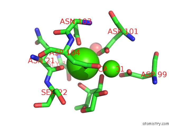
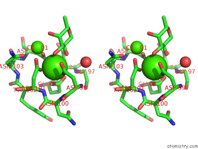
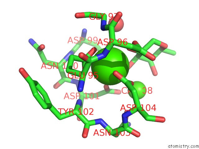
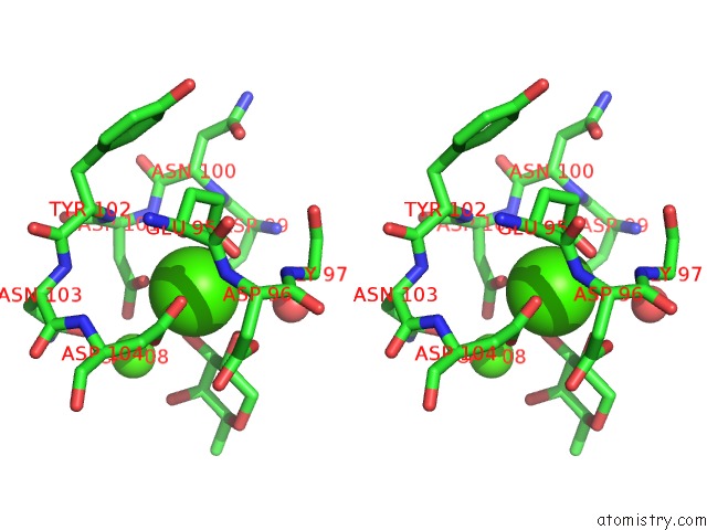
Calcium binding site 2 out of 8 in 6r35
Go back to
Calcium binding site 2 out
of 8 in the Structure of the Lecb Lectin From Pseudomonas Aeruginosa Strain PAO1 in Complex with Lewis X Tetrasaccharide

Mono view
Stereo pair view
Mono view
Stereo pair view
A full contact list of Calcium with other atoms in the Ca binding
site number 2 of Structure of the Lecb Lectin From Pseudomonas Aeruginosa Strain PAO1 in Complex with Lewis X Tetrasaccharide within 5.0Å range:
|
Calcium binding site 3 out of 8 in 6r35
Go back to
Calcium binding site 3 out
of 8 in the Structure of the Lecb Lectin From Pseudomonas Aeruginosa Strain PAO1 in Complex with Lewis X Tetrasaccharide
Mono view

Stereo pair view
Mono view
Stereo pair view
A full contact list of Calcium with other atoms in the Ca binding
site number 3 of Structure of the Lecb Lectin From Pseudomonas Aeruginosa Strain PAO1 in Complex with Lewis X Tetrasaccharide within 5.0Å range:
|
Calcium binding site 4 out of 8 in 6r35
Go back to
Calcium binding site 4 out
of 8 in the Structure of the Lecb Lectin From Pseudomonas Aeruginosa Strain PAO1 in Complex with Lewis X Tetrasaccharide

Mono view

Stereo pair view
Mono view
Stereo pair view
A full contact list of Calcium with other atoms in the Ca binding
site number 4 of Structure of the Lecb Lectin From Pseudomonas Aeruginosa Strain PAO1 in Complex with Lewis X Tetrasaccharide within 5.0Å range:
|
Calcium binding site 5 out of 8 in 6r35
Go back to
Calcium binding site 5 out
of 8 in the Structure of the Lecb Lectin From Pseudomonas Aeruginosa Strain PAO1 in Complex with Lewis X Tetrasaccharide

Mono view
Stereo pair view
Mono view
Stereo pair view
A full contact list of Calcium with other atoms in the Ca binding
site number 5 of Structure of the Lecb Lectin From Pseudomonas Aeruginosa Strain PAO1 in Complex with Lewis X Tetrasaccharide within 5.0Å range:
|
Calcium binding site 6 out of 8 in 6r35
Go back to
Calcium binding site 6 out
of 8 in the Structure of the Lecb Lectin From Pseudomonas Aeruginosa Strain PAO1 in Complex with Lewis X Tetrasaccharide
Mono view

Stereo pair view
Mono view
Stereo pair view
A full contact list of Calcium with other atoms in the Ca binding
site number 6 of Structure of the Lecb Lectin From Pseudomonas Aeruginosa Strain PAO1 in Complex with Lewis X Tetrasaccharide within 5.0Å range:
|
Calcium binding site 7 out of 8 in 6r35
Go back to
Calcium binding site 7 out
of 8 in the Structure of the Lecb Lectin From Pseudomonas Aeruginosa Strain PAO1 in Complex with Lewis X Tetrasaccharide
Mono view

Stereo pair view
Mono view
Stereo pair view
A full contact list of Calcium with other atoms in the Ca binding
site number 7 of Structure of the Lecb Lectin From Pseudomonas Aeruginosa Strain PAO1 in Complex with Lewis X Tetrasaccharide within 5.0Å range:
|
Calcium binding site 8 out of 8 in 6r35
Go back to
Calcium binding site 8 out
of 8 in the Structure of the Lecb Lectin From Pseudomonas Aeruginosa Strain PAO1 in Complex with Lewis X Tetrasaccharide

Mono view
Stereo pair view
Mono view
Stereo pair view
A full contact list of Calcium with other atoms in the Ca binding
site number 8 of Structure of the Lecb Lectin From Pseudomonas Aeruginosa Strain PAO1 in Complex with Lewis X Tetrasaccharide within 5.0Å range:
|
Reference:
M.Lepsik,
R.Sommer,
S.Kuhaudomlarp,
M.Lelimousin,
E.Paci,
A.Varrot,
A.Titz,
A.Imberty.
Induction of Rare Conformation of Oligosaccharide By Binding to Calcium-Dependent Bacterial Lectin: X-Ray Crystallography and Modelling Study. Eur.J.Med.Chem. V. 177 212 2019.
ISSN: ISSN 0223-5234
PubMed: 31146126
DOI: 10.1016/J.EJMECH.2019.05.049
Page generated: Tue Jul 16 14:14:26 2024
ISSN: ISSN 0223-5234
PubMed: 31146126
DOI: 10.1016/J.EJMECH.2019.05.049
Last articles
Zn in 9MJ5Zn in 9HNW
Zn in 9G0L
Zn in 9FNE
Zn in 9DZN
Zn in 9E0I
Zn in 9D32
Zn in 9DAK
Zn in 8ZXC
Zn in 8ZUF