Calcium »
PDB 6r7u-6ry5 »
6rkj »
Calcium in PDB 6rkj: The Crystal Structure of Abne, An Arabino-Oligosaccharide Binding Protein, in Complex with Arabinooctaose
Protein crystallography data
The structure of The Crystal Structure of Abne, An Arabino-Oligosaccharide Binding Protein, in Complex with Arabinooctaose, PDB code: 6rkj
was solved by
S.Lansky,
R.Salama,
Y.Shoham,
G.Shoham,
with X-Ray Crystallography technique. A brief refinement statistics is given in the table below:
| Resolution Low / High (Å) | 34.91 / 1.60 |
| Space group | P 21 21 21 |
| Cell size a, b, c (Å), α, β, γ (°) | 39.477, 95.608, 120.076, 90.00, 90.00, 90.00 |
| R / Rfree (%) | 15.9 / 18.8 |
Calcium Binding Sites:
The binding sites of Calcium atom in the The Crystal Structure of Abne, An Arabino-Oligosaccharide Binding Protein, in Complex with Arabinooctaose
(pdb code 6rkj). This binding sites where shown within
5.0 Angstroms radius around Calcium atom.
In total only one binding site of Calcium was determined in the The Crystal Structure of Abne, An Arabino-Oligosaccharide Binding Protein, in Complex with Arabinooctaose, PDB code: 6rkj:
In total only one binding site of Calcium was determined in the The Crystal Structure of Abne, An Arabino-Oligosaccharide Binding Protein, in Complex with Arabinooctaose, PDB code: 6rkj:
Calcium binding site 1 out of 1 in 6rkj
Go back to
Calcium binding site 1 out
of 1 in the The Crystal Structure of Abne, An Arabino-Oligosaccharide Binding Protein, in Complex with Arabinooctaose
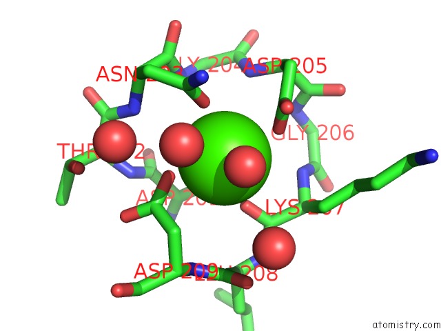
Mono view
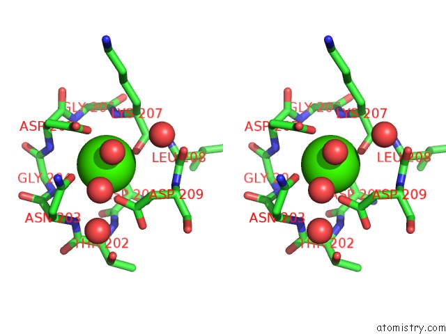
Stereo pair view
Mono view
Stereo pair view
A full contact list of Calcium with other atoms in the Ca binding
site number 1 of The Crystal Structure of Abne, An Arabino-Oligosaccharide Binding Protein, in Complex with Arabinooctaose within 5.0Å range:
|
Reference:
S.Lansky,
R.Salama,
S.Shulami,
N.Lavid,
S.Sen,
I.Schapiro,
Y.Shoham,
G.Shoham.
Carbohydrate-Binding Capability and Functional Conformational Changes of Abne, An Arabino-Oligosaccharide Binding Protein. J.Mol.Biol. 2020.
ISSN: ESSN 1089-8638
PubMed: 32067952
DOI: 10.1016/J.JMB.2020.01.041
Page generated: Tue Jul 16 14:23:47 2024
ISSN: ESSN 1089-8638
PubMed: 32067952
DOI: 10.1016/J.JMB.2020.01.041
Last articles
Zn in 9J0NZn in 9J0O
Zn in 9J0P
Zn in 9FJX
Zn in 9EKB
Zn in 9C0F
Zn in 9CAH
Zn in 9CH0
Zn in 9CH3
Zn in 9CH1