Calcium »
PDB 6r7u-6ry5 »
6ry1 »
Calcium in PDB 6ry1: Crystal Structure of DFG5 From Chaetomium Thermophilum in Complex with Mannose
Enzymatic activity of Crystal Structure of DFG5 From Chaetomium Thermophilum in Complex with Mannose
All present enzymatic activity of Crystal Structure of DFG5 From Chaetomium Thermophilum in Complex with Mannose:
3.2.1.101;
3.2.1.101;
Protein crystallography data
The structure of Crystal Structure of DFG5 From Chaetomium Thermophilum in Complex with Mannose, PDB code: 6ry1
was solved by
L.-O.Essen,
M.S.Vogt,
with X-Ray Crystallography technique. A brief refinement statistics is given in the table below:
| Resolution Low / High (Å) | 41.93 / 1.30 |
| Space group | C 1 2 1 |
| Cell size a, b, c (Å), α, β, γ (°) | 83.855, 55.022, 80.458, 90.00, 90.45, 90.00 |
| R / Rfree (%) | 9.6 / 12.5 |
Calcium Binding Sites:
The binding sites of Calcium atom in the Crystal Structure of DFG5 From Chaetomium Thermophilum in Complex with Mannose
(pdb code 6ry1). This binding sites where shown within
5.0 Angstroms radius around Calcium atom.
In total 2 binding sites of Calcium where determined in the Crystal Structure of DFG5 From Chaetomium Thermophilum in Complex with Mannose, PDB code: 6ry1:
Jump to Calcium binding site number: 1; 2;
In total 2 binding sites of Calcium where determined in the Crystal Structure of DFG5 From Chaetomium Thermophilum in Complex with Mannose, PDB code: 6ry1:
Jump to Calcium binding site number: 1; 2;
Calcium binding site 1 out of 2 in 6ry1
Go back to
Calcium binding site 1 out
of 2 in the Crystal Structure of DFG5 From Chaetomium Thermophilum in Complex with Mannose
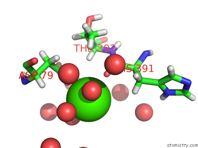
Mono view
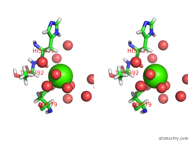
Stereo pair view
Mono view
Stereo pair view
A full contact list of Calcium with other atoms in the Ca binding
site number 1 of Crystal Structure of DFG5 From Chaetomium Thermophilum in Complex with Mannose within 5.0Å range:
|
Calcium binding site 2 out of 2 in 6ry1
Go back to
Calcium binding site 2 out
of 2 in the Crystal Structure of DFG5 From Chaetomium Thermophilum in Complex with Mannose
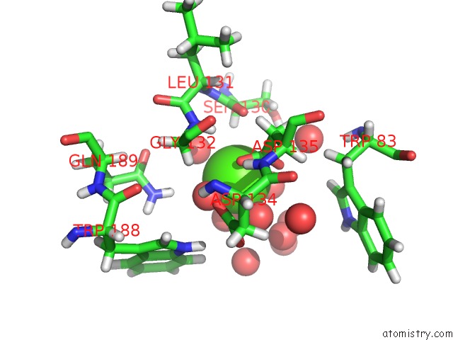
Mono view
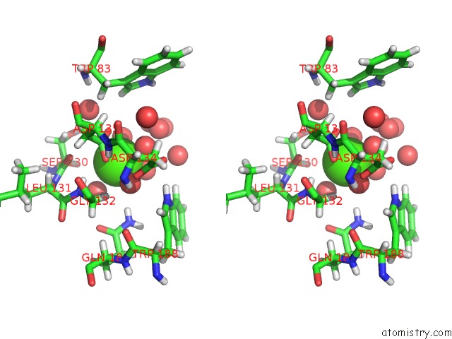
Stereo pair view
Mono view
Stereo pair view
A full contact list of Calcium with other atoms in the Ca binding
site number 2 of Crystal Structure of DFG5 From Chaetomium Thermophilum in Complex with Mannose within 5.0Å range:
|
Reference:
M.S.Vogt,
G.F.Schmitz,
D.Varon Silva,
H.U.Mosch,
L.O.Essen.
Structural Base For the Transfer of Gpi-Anchored Glycoproteins Into Fungal Cell Walls. Proc.Natl.Acad.Sci.Usa V. 117 22061 2020.
ISSN: ESSN 1091-6490
PubMed: 32839341
DOI: 10.1073/PNAS.2010661117
Page generated: Tue Jul 16 14:27:06 2024
ISSN: ESSN 1091-6490
PubMed: 32839341
DOI: 10.1073/PNAS.2010661117
Last articles
Zn in 9J0NZn in 9J0O
Zn in 9J0P
Zn in 9FJX
Zn in 9EKB
Zn in 9C0F
Zn in 9CAH
Zn in 9CH0
Zn in 9CH3
Zn in 9CH1