Calcium »
PDB 6ry6-6sav »
6ryn »
Calcium in PDB 6ryn: Structure of Conglutinin Carbohydrate Recognition Domain with Glcnac- Alpha-1-Phosphate Bound
Protein crystallography data
The structure of Structure of Conglutinin Carbohydrate Recognition Domain with Glcnac- Alpha-1-Phosphate Bound, PDB code: 6ryn
was solved by
A.K.Shrive,
T.J.Greenhough,
with X-Ray Crystallography technique. A brief refinement statistics is given in the table below:
| Resolution Low / High (Å) | 50.00 / 1.00 |
| Space group | P 43 |
| Cell size a, b, c (Å), α, β, γ (°) | 50.278, 50.278, 52.192, 90.00, 90.00, 90.00 |
| R / Rfree (%) | 12.4 / 14 |
Calcium Binding Sites:
The binding sites of Calcium atom in the Structure of Conglutinin Carbohydrate Recognition Domain with Glcnac- Alpha-1-Phosphate Bound
(pdb code 6ryn). This binding sites where shown within
5.0 Angstroms radius around Calcium atom.
In total 2 binding sites of Calcium where determined in the Structure of Conglutinin Carbohydrate Recognition Domain with Glcnac- Alpha-1-Phosphate Bound, PDB code: 6ryn:
Jump to Calcium binding site number: 1; 2;
In total 2 binding sites of Calcium where determined in the Structure of Conglutinin Carbohydrate Recognition Domain with Glcnac- Alpha-1-Phosphate Bound, PDB code: 6ryn:
Jump to Calcium binding site number: 1; 2;
Calcium binding site 1 out of 2 in 6ryn
Go back to
Calcium binding site 1 out
of 2 in the Structure of Conglutinin Carbohydrate Recognition Domain with Glcnac- Alpha-1-Phosphate Bound
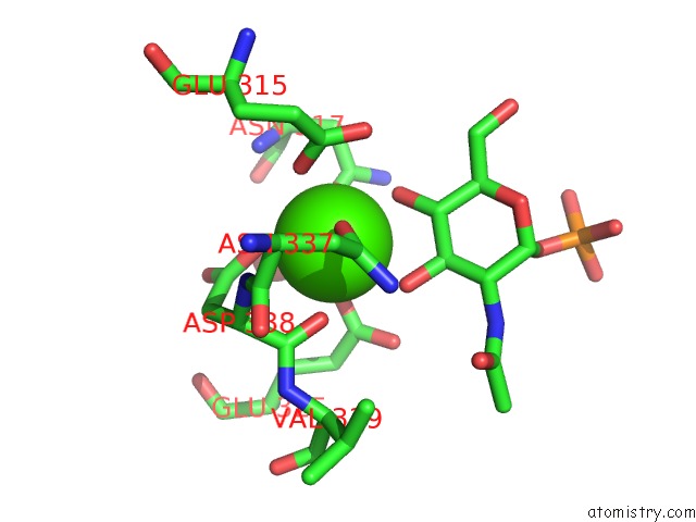
Mono view
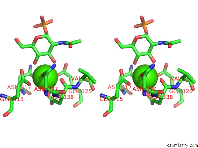
Stereo pair view
Mono view
Stereo pair view
A full contact list of Calcium with other atoms in the Ca binding
site number 1 of Structure of Conglutinin Carbohydrate Recognition Domain with Glcnac- Alpha-1-Phosphate Bound within 5.0Å range:
|
Calcium binding site 2 out of 2 in 6ryn
Go back to
Calcium binding site 2 out
of 2 in the Structure of Conglutinin Carbohydrate Recognition Domain with Glcnac- Alpha-1-Phosphate Bound
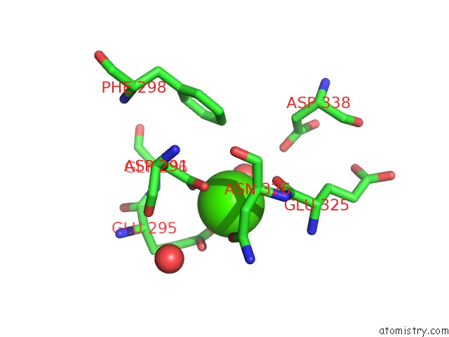
Mono view
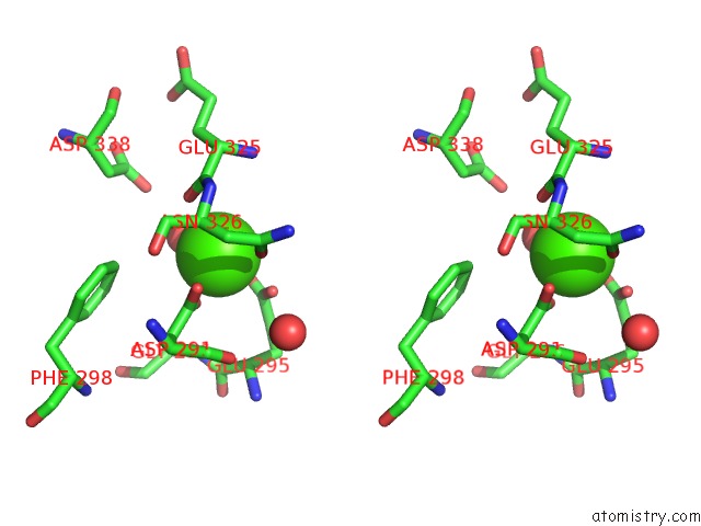
Stereo pair view
Mono view
Stereo pair view
A full contact list of Calcium with other atoms in the Ca binding
site number 2 of Structure of Conglutinin Carbohydrate Recognition Domain with Glcnac- Alpha-1-Phosphate Bound within 5.0Å range:
|
Reference:
J.M.Paterson,
A.J.Shaw,
I.Burns,
A.W.Dodds,
A.Prasad,
K.B.Reid,
T.J.Greenhough,
A.K.Shrive.
Atomic-Resolution Crystal Structures of the Immune Protein Conglutinin From Cow Reveal Specific Interactions of Its Binding Site Withn-Acetylglucosamine. J.Biol.Chem. V. 294 17155 2019.
ISSN: ESSN 1083-351X
PubMed: 31562242
DOI: 10.1074/JBC.RA119.010271
Page generated: Tue Jul 16 14:28:47 2024
ISSN: ESSN 1083-351X
PubMed: 31562242
DOI: 10.1074/JBC.RA119.010271
Last articles
Zn in 9J0NZn in 9J0O
Zn in 9J0P
Zn in 9FJX
Zn in 9EKB
Zn in 9C0F
Zn in 9CAH
Zn in 9CH0
Zn in 9CH3
Zn in 9CH1