Calcium »
PDB 6sbk-6sux »
6sit »
Calcium in PDB 6sit: Pseudo-Atomic Crystal Structure of the Desmoglein 2 - Human Adenovirus Serotype 3 Fibre Knob Complex
Protein crystallography data
The structure of Pseudo-Atomic Crystal Structure of the Desmoglein 2 - Human Adenovirus Serotype 3 Fibre Knob Complex, PDB code: 6sit
was solved by
W.P.Burmeister,
P.Fender,
E.Vassal-Stermann,
with X-Ray Crystallography technique. A brief refinement statistics is given in the table below:
| Resolution Low / High (Å) | 46.50 / 4.50 |
| Space group | I 21 3 |
| Cell size a, b, c (Å), α, β, γ (°) | 146.530, 146.530, 146.530, 90.00, 90.00, 90.00 |
| R / Rfree (%) | 36.7 / 37 |
Calcium Binding Sites:
The binding sites of Calcium atom in the Pseudo-Atomic Crystal Structure of the Desmoglein 2 - Human Adenovirus Serotype 3 Fibre Knob Complex
(pdb code 6sit). This binding sites where shown within
5.0 Angstroms radius around Calcium atom.
In total 6 binding sites of Calcium where determined in the Pseudo-Atomic Crystal Structure of the Desmoglein 2 - Human Adenovirus Serotype 3 Fibre Knob Complex, PDB code: 6sit:
Jump to Calcium binding site number: 1; 2; 3; 4; 5; 6;
In total 6 binding sites of Calcium where determined in the Pseudo-Atomic Crystal Structure of the Desmoglein 2 - Human Adenovirus Serotype 3 Fibre Knob Complex, PDB code: 6sit:
Jump to Calcium binding site number: 1; 2; 3; 4; 5; 6;
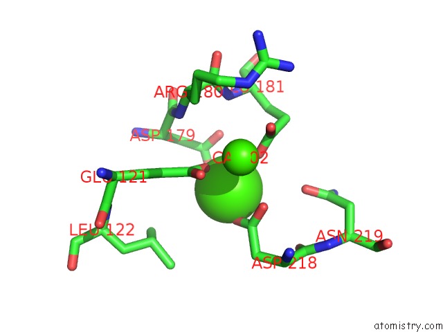
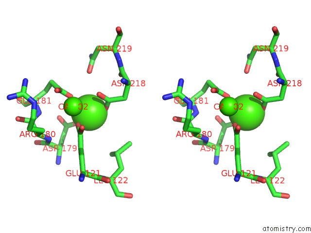
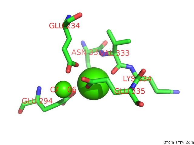
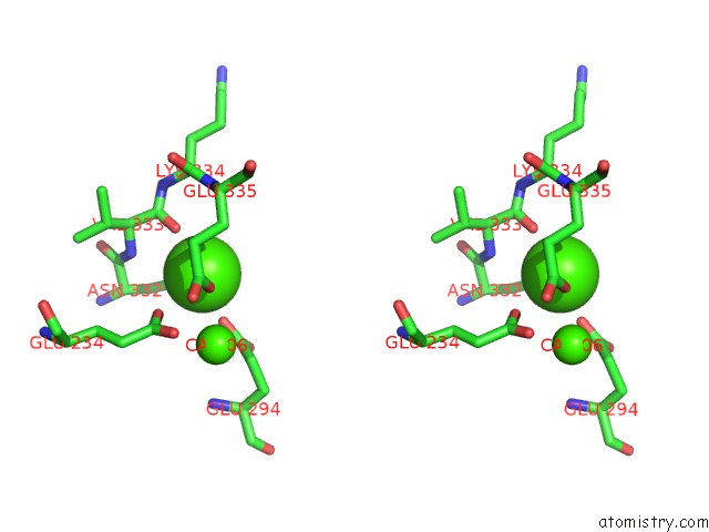
Calcium binding site 1 out of 6 in 6sit
Go back to
Calcium binding site 1 out
of 6 in the Pseudo-Atomic Crystal Structure of the Desmoglein 2 - Human Adenovirus Serotype 3 Fibre Knob Complex
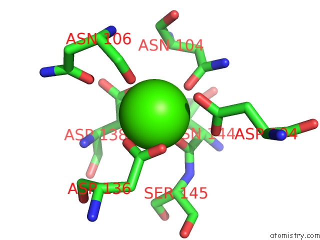
Mono view
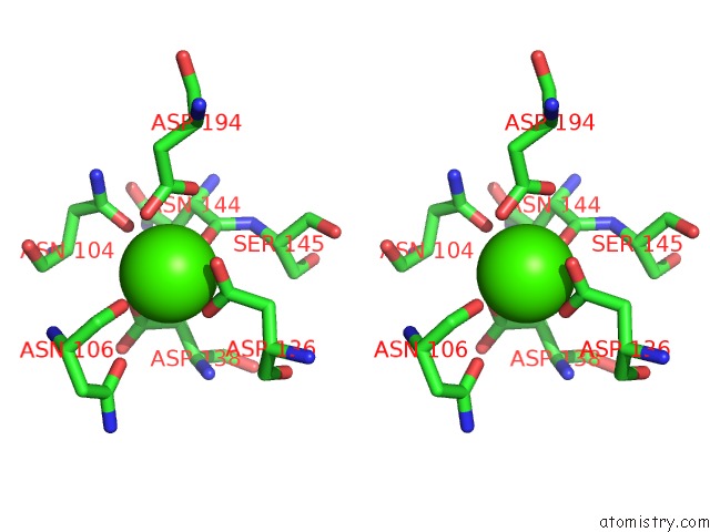
Stereo pair view
Mono view
Stereo pair view
A full contact list of Calcium with other atoms in the Ca binding
site number 1 of Pseudo-Atomic Crystal Structure of the Desmoglein 2 - Human Adenovirus Serotype 3 Fibre Knob Complex within 5.0Å range:
|
Calcium binding site 2 out of 6 in 6sit
Go back to
Calcium binding site 2 out
of 6 in the Pseudo-Atomic Crystal Structure of the Desmoglein 2 - Human Adenovirus Serotype 3 Fibre Knob Complex
Mono view
Stereo pair view
Mono view
Stereo pair view
A full contact list of Calcium with other atoms in the Ca binding
site number 2 of Pseudo-Atomic Crystal Structure of the Desmoglein 2 - Human Adenovirus Serotype 3 Fibre Knob Complex within 5.0Å range:
|
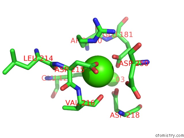
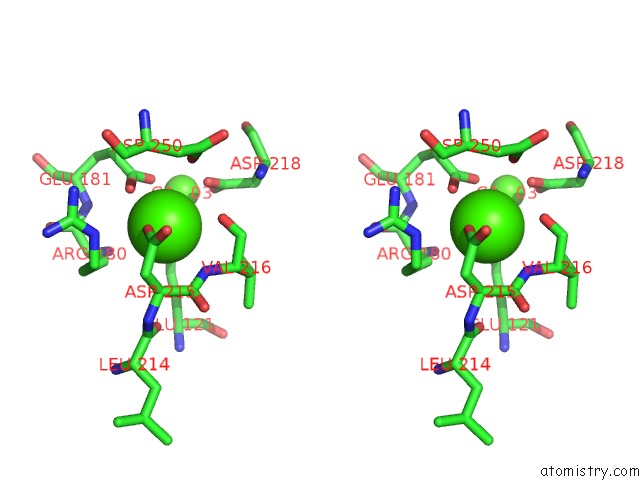
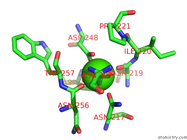
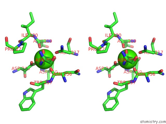
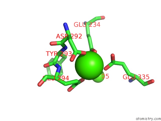
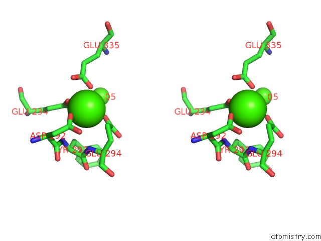
Calcium binding site 3 out of 6 in 6sit
Go back to
Calcium binding site 3 out
of 6 in the Pseudo-Atomic Crystal Structure of the Desmoglein 2 - Human Adenovirus Serotype 3 Fibre Knob Complex
Mono view
Stereo pair view
Mono view
Stereo pair view
A full contact list of Calcium with other atoms in the Ca binding
site number 3 of Pseudo-Atomic Crystal Structure of the Desmoglein 2 - Human Adenovirus Serotype 3 Fibre Knob Complex within 5.0Å range:
|
Calcium binding site 4 out of 6 in 6sit
Go back to
Calcium binding site 4 out
of 6 in the Pseudo-Atomic Crystal Structure of the Desmoglein 2 - Human Adenovirus Serotype 3 Fibre Knob Complex
Mono view
Stereo pair view
Mono view
Stereo pair view
A full contact list of Calcium with other atoms in the Ca binding
site number 4 of Pseudo-Atomic Crystal Structure of the Desmoglein 2 - Human Adenovirus Serotype 3 Fibre Knob Complex within 5.0Å range:
|
Calcium binding site 5 out of 6 in 6sit
Go back to
Calcium binding site 5 out
of 6 in the Pseudo-Atomic Crystal Structure of the Desmoglein 2 - Human Adenovirus Serotype 3 Fibre Knob Complex
Mono view
Stereo pair view
Mono view
Stereo pair view
A full contact list of Calcium with other atoms in the Ca binding
site number 5 of Pseudo-Atomic Crystal Structure of the Desmoglein 2 - Human Adenovirus Serotype 3 Fibre Knob Complex within 5.0Å range:
|
Calcium binding site 6 out of 6 in 6sit
Go back to
Calcium binding site 6 out
of 6 in the Pseudo-Atomic Crystal Structure of the Desmoglein 2 - Human Adenovirus Serotype 3 Fibre Knob Complex
Mono view
Stereo pair view
Mono view
Stereo pair view
A full contact list of Calcium with other atoms in the Ca binding
site number 6 of Pseudo-Atomic Crystal Structure of the Desmoglein 2 - Human Adenovirus Serotype 3 Fibre Knob Complex within 5.0Å range:
|
Reference:
E.Vassal-Stermann,
S.Hutin,
P.Fender,
W.P.Burmeister.
Intermediate-Resolution Crystal Structure of the Human Adenovirus B Serotype 3 Fibre Knob in Complex with the EC2-EC3 Fragment of Desmoglein 2. Acta Crystallogr.,Sect.F V. 75 750 2019.
ISSN: ESSN 2053-230X
PubMed: 31797817
DOI: 10.1107/S2053230X19015784
Page generated: Wed Jul 9 17:50:15 2025
ISSN: ESSN 2053-230X
PubMed: 31797817
DOI: 10.1107/S2053230X19015784
Last articles
F in 7MOGF in 7MOO
F in 7MML
F in 7MMI
F in 7MMK
F in 7MMJ
F in 7MMG
F in 7MMF
F in 7MMH
F in 7MMA