Calcium »
PDB 6suw-6t6v »
6svn »
Calcium in PDB 6svn: Reference Structure of Bovine Trypsin (Even Frames of Crystal X28)
Enzymatic activity of Reference Structure of Bovine Trypsin (Even Frames of Crystal X28)
All present enzymatic activity of Reference Structure of Bovine Trypsin (Even Frames of Crystal X28):
3.4.21.4;
3.4.21.4;
Protein crystallography data
The structure of Reference Structure of Bovine Trypsin (Even Frames of Crystal X28), PDB code: 6svn
was solved by
V.Ahlberg Gagner,
I.Lundholm,
M.J.Garcia-Bonete,
H.Rodilla,
R.Friedman,
V.Zhaunerchyk,
G.Bourenkov,
T.Schneider,
J.Stake,
G.Katona,
with X-Ray Crystallography technique. A brief refinement statistics is given in the table below:
| Resolution Low / High (Å) | 42.89 / 1.16 |
| Space group | P 21 21 21 |
| Cell size a, b, c (Å), α, β, γ (°) | 53.410, 56.860, 65.190, 90.00, 90.00, 90.00 |
| R / Rfree (%) | 13 / 17.1 |
Calcium Binding Sites:
The binding sites of Calcium atom in the Reference Structure of Bovine Trypsin (Even Frames of Crystal X28)
(pdb code 6svn). This binding sites where shown within
5.0 Angstroms radius around Calcium atom.
In total only one binding site of Calcium was determined in the Reference Structure of Bovine Trypsin (Even Frames of Crystal X28), PDB code: 6svn:
In total only one binding site of Calcium was determined in the Reference Structure of Bovine Trypsin (Even Frames of Crystal X28), PDB code: 6svn:
Calcium binding site 1 out of 1 in 6svn
Go back to
Calcium binding site 1 out
of 1 in the Reference Structure of Bovine Trypsin (Even Frames of Crystal X28)
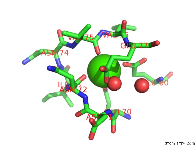
Mono view
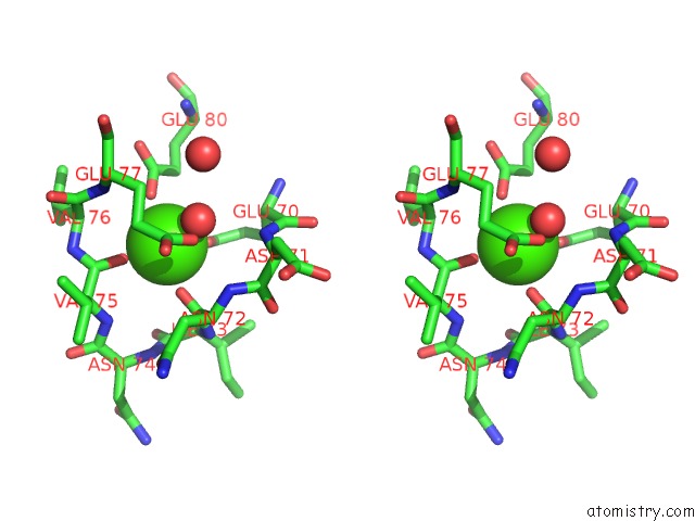
Stereo pair view
Mono view
Stereo pair view
A full contact list of Calcium with other atoms in the Ca binding
site number 1 of Reference Structure of Bovine Trypsin (Even Frames of Crystal X28) within 5.0Å range:
|
Reference:
V.Ahlberg Gagner,
I.Lundholm,
M.J.Garcia-Bonete,
H.Rodilla,
R.Friedman,
V.Zhaunerchyk,
G.Bourenkov,
T.Schneider,
J.Stake,
G.Katona.
Clustering of Atomic Displacement Parameters in Bovine Trypsin Reveals A Distributed Lattice of Atoms with Shared Chemical Properties Sci Rep 2020.
ISSN: ESSN 2045-2322
Page generated: Tue Jul 16 14:59:51 2024
ISSN: ESSN 2045-2322
Last articles
Zn in 9JYWZn in 9IR4
Zn in 9IR3
Zn in 9GMX
Zn in 9GMW
Zn in 9JEJ
Zn in 9ERF
Zn in 9ERE
Zn in 9EGV
Zn in 9EGW