Calcium »
PDB 6suw-6t6v »
6t0p »
Calcium in PDB 6t0p: Cationic Trypsin in Complex with A D-Phe-Pro-2-Aminopyridine Derivative
Enzymatic activity of Cationic Trypsin in Complex with A D-Phe-Pro-2-Aminopyridine Derivative
All present enzymatic activity of Cationic Trypsin in Complex with A D-Phe-Pro-2-Aminopyridine Derivative:
3.4.21.4;
3.4.21.4;
Protein crystallography data
The structure of Cationic Trypsin in Complex with A D-Phe-Pro-2-Aminopyridine Derivative, PDB code: 6t0p
was solved by
K.Ngo,
A.Heine,
G.Klebe,
with X-Ray Crystallography technique. A brief refinement statistics is given in the table below:
| Resolution Low / High (Å) | 39.31 / 1.19 |
| Space group | P 21 21 21 |
| Cell size a, b, c (Å), α, β, γ (°) | 54.568, 56.684, 66.647, 90.00, 90.00, 90.00 |
| R / Rfree (%) | 12.3 / 13.8 |
Calcium Binding Sites:
The binding sites of Calcium atom in the Cationic Trypsin in Complex with A D-Phe-Pro-2-Aminopyridine Derivative
(pdb code 6t0p). This binding sites where shown within
5.0 Angstroms radius around Calcium atom.
In total only one binding site of Calcium was determined in the Cationic Trypsin in Complex with A D-Phe-Pro-2-Aminopyridine Derivative, PDB code: 6t0p:
In total only one binding site of Calcium was determined in the Cationic Trypsin in Complex with A D-Phe-Pro-2-Aminopyridine Derivative, PDB code: 6t0p:
Calcium binding site 1 out of 1 in 6t0p
Go back to
Calcium binding site 1 out
of 1 in the Cationic Trypsin in Complex with A D-Phe-Pro-2-Aminopyridine Derivative
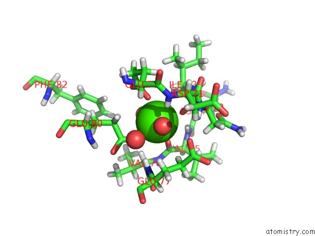
Mono view
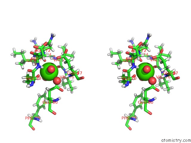
Stereo pair view
Mono view
Stereo pair view
A full contact list of Calcium with other atoms in the Ca binding
site number 1 of Cationic Trypsin in Complex with A D-Phe-Pro-2-Aminopyridine Derivative within 5.0Å range:
|
Reference:
K.Ngo,
C.Collins-Kautz,
S.Gerstenecker,
B.Wagner,
A.Heine,
G.Klebe.
Protein-Induced Change in Ligand Protonation During Trypsin and Thrombin Binding: Hint on Differences in Selectivity Determinants of Both Proteins? J.Med.Chem. V. 63 3274 2020.
ISSN: ISSN 0022-2623
PubMed: 32011145
DOI: 10.1021/ACS.JMEDCHEM.9B02061
Page generated: Tue Jul 16 15:04:15 2024
ISSN: ISSN 0022-2623
PubMed: 32011145
DOI: 10.1021/ACS.JMEDCHEM.9B02061
Last articles
Zn in 9JYWZn in 9IR4
Zn in 9IR3
Zn in 9GMX
Zn in 9GMW
Zn in 9JEJ
Zn in 9ERF
Zn in 9ERE
Zn in 9EGV
Zn in 9EGW