Calcium in PDB 6t8f: Crystal Structure of Mutant Xylose Isomerase (V270A/A273G) From Piromyces E2 Grown in Yeast, in Complex with Xylose

Enzymatic activity of Crystal Structure of Mutant Xylose Isomerase (V270A/A273G) From Piromyces E2 Grown in Yeast, in Complex with Xylose

All present enzymatic activity of Crystal Structure of Mutant Xylose Isomerase (V270A/A273G) From Piromyces E2 Grown in Yeast, in Complex with Xylose:
5.3.1.5;

Protein crystallography data

The structure of Crystal Structure of Mutant Xylose Isomerase (V270A/A273G) From Piromyces E2 Grown in Yeast, in Complex with Xylose, PDB code: 6t8f was solved by H.J.Rozeboom, D.B.Janssen, with X-Ray Crystallography technique. A brief refinement statistics is given in the table below:

Resolution Low / High (Å) 46.60 / 2.00
Space group P 1
Cell size a, b, c (Å), α, β, γ (°) 78.598, 79.372, 91.982, 115.46, 89.98, 117.13
R / Rfree (%) 14.9 / 18.5

Calcium Binding Sites:

The binding sites of Calcium atom in the Crystal Structure of Mutant Xylose Isomerase (V270A/A273G) From Piromyces E2 Grown in Yeast, in Complex with Xylose (pdb code 6t8f). This binding sites where shown within 5.0 Angstroms radius around Calcium atom.
In total 8 binding sites of Calcium where determined in the Crystal Structure of Mutant Xylose Isomerase (V270A/A273G) From Piromyces E2 Grown in Yeast, in Complex with Xylose, PDB code: 6t8f:
Jump to Calcium binding site number: 1; 2; 3; 4; 5; 6; 7; 8;

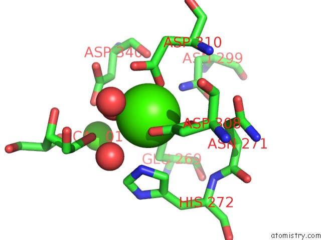
Calcium binding site 1 out of 8 in 6t8f

Go back to Calcium Binding Sites List in 6t8f
Calcium binding site 1 out of 8 in the Crystal Structure of Mutant Xylose Isomerase (V270A/A273G) From Piromyces E2 Grown in Yeast, in Complex with Xylose


Mono view


Stereo pair view

A full contact list of Calcium with other atoms in the Ca binding site number 1 of Crystal Structure of Mutant Xylose Isomerase (V270A/A273G) From Piromyces E2 Grown in Yeast, in Complex with Xylose within 5.0Å range:
probe atom residue distance (Å) B Occ
A:Ca501

b:16.5
occ:1.00
OD2 A:ASP297 2.1 19.7 1.0
OE2 A:GLU233 2.1 19.9 1.0
OE1 A:GLU269 2.1 21.7 1.0
O2 A:XLS503 2.2 29.7 1.0
OD2 A:ASP340 2.2 20.9 1.0
O4 A:XLS503 2.3 29.4 1.0
CD A:GLU233 2.9 18.5 1.0
OE1 A:GLU233 3.1 21.1 1.0
C2 A:XLS503 3.2 33.8 1.0
CG A:ASP297 3.2 19.9 1.0
C4 A:XLS503 3.3 32.5 1.0
CG A:ASP340 3.3 19.1 1.0
CD A:GLU269 3.3 19.3 1.0
C3 A:XLS503 3.5 34.6 1.0
O3 A:XLS503 3.6 36.9 1.0
CB A:ASP297 3.8 17.9 1.0
CB A:ASP340 3.8 17.4 1.0
O A:HOH614 3.9 20.3 1.0
CE1 A:HIS272 3.9 17.5 1.0
CG A:GLU269 4.1 18.6 1.0
CB A:GLU269 4.1 17.8 1.0
OE2 A:GLU269 4.3 20.2 1.0
CG A:GLU233 4.3 18.4 1.0
OD1 A:ASP297 4.3 20.1 1.0
OD1 A:ASP340 4.4 19.7 1.0
NE2 A:HIS272 4.5 17.9 1.0
ND1 A:HIS272 4.6 16.1 1.0
C1 A:XLS503 4.6 32.9 1.0
ND2 A:ASN267 4.6 16.4 1.0
C5 A:XLS503 4.7 32.2 1.0
CA A:CA502 4.8 21.0 1.0

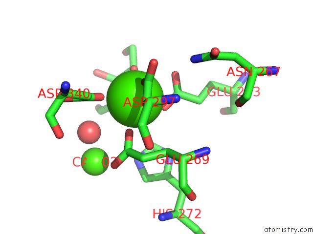
Calcium binding site 2 out of 8 in 6t8f

Go back to Calcium Binding Sites List in 6t8f
Calcium binding site 2 out of 8 in the Crystal Structure of Mutant Xylose Isomerase (V270A/A273G) From Piromyces E2 Grown in Yeast, in Complex with Xylose


Mono view


Stereo pair view

A full contact list of Calcium with other atoms in the Ca binding site number 2 of Crystal Structure of Mutant Xylose Isomerase (V270A/A273G) From Piromyces E2 Grown in Yeast, in Complex with Xylose within 5.0Å range:
probe atom residue distance (Å) B Occ
A:Ca502

b:21.0
occ:1.00
OD1 A:ASP310 1.9 22.0 1.0
OD2 A:ASP308 1.9 33.0 1.0
O A:HOH614 2.0 20.3 1.0
OE2 A:GLU269 2.0 20.2 1.0
CG A:ASP308 2.6 29.8 1.0
OD1 A:ASP308 2.7 35.7 1.0
CG A:ASP310 2.9 21.0 1.0
CD A:GLU269 2.9 19.3 1.0
NE2 A:HIS272 3.1 17.9 1.0
OD2 A:ASP310 3.1 21.4 1.0
OE1 A:GLU269 3.2 21.7 1.0
O A:HOH675 3.5 25.4 1.0
CD2 A:HIS272 3.6 16.5 1.0
O2 A:XLS503 3.7 29.7 1.0
ND2 A:ASN299 3.8 13.6 1.0
O A:HOH898 4.0 37.7 1.0
CB A:ASP308 4.1 25.6 1.0
CE1 A:HIS272 4.1 17.5 1.0
CG A:GLU269 4.3 18.6 1.0
OD2 A:ASP340 4.3 20.9 1.0
CB A:ASP310 4.3 20.0 1.0
CG A:ASP340 4.5 19.1 1.0
OD1 A:ASP340 4.6 19.7 1.0
CB A:ASN271 4.6 17.2 1.0
O1 A:XLS503 4.7 33.8 1.0
O A:ASP340 4.7 15.6 1.0
C2 A:XLS503 4.8 33.8 1.0
CA A:ASP310 4.8 18.6 1.0
CA A:CA501 4.8 16.5 1.0
CG A:HIS272 4.8 16.7 1.0
C1 A:XLS503 4.9 32.9 1.0
N A:ASP310 5.0 17.3 1.0

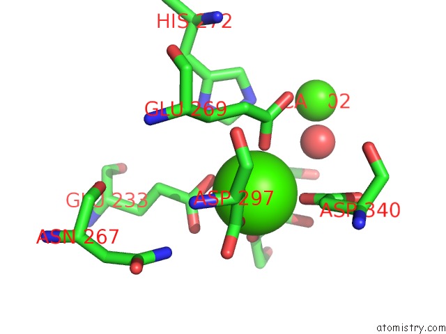
Calcium binding site 3 out of 8 in 6t8f

Go back to Calcium Binding Sites List in 6t8f
Calcium binding site 3 out of 8 in the Crystal Structure of Mutant Xylose Isomerase (V270A/A273G) From Piromyces E2 Grown in Yeast, in Complex with Xylose


Mono view


Stereo pair view

A full contact list of Calcium with other atoms in the Ca binding site number 3 of Crystal Structure of Mutant Xylose Isomerase (V270A/A273G) From Piromyces E2 Grown in Yeast, in Complex with Xylose within 5.0Å range:
probe atom residue distance (Å) B Occ
B:Ca501

b:16.5
occ:1.00
OE2 B:GLU233 2.1 19.4 1.0
OD2 B:ASP297 2.1 22.1 1.0
O2 B:XLS503 2.1 34.5 1.0
OE1 B:GLU269 2.1 23.9 1.0
OD2 B:ASP340 2.2 19.9 1.0
O4 B:XLS503 2.4 32.3 1.0
CD B:GLU233 2.9 18.4 1.0
OE1 B:GLU233 3.1 18.3 1.0
CG B:ASP297 3.2 19.1 1.0
C2 B:XLS503 3.3 38.2 1.0
CG B:ASP340 3.3 19.6 1.0
CD B:GLU269 3.3 22.4 1.0
C4 B:XLS503 3.4 35.3 1.0
CB B:ASP297 3.7 17.6 1.0
O B:HOH637 3.7 26.3 1.0
CB B:ASP340 3.8 18.0 1.0
C3 B:XLS503 3.8 38.0 1.0
CE1 B:HIS272 4.0 22.3 1.0
CG B:GLU269 4.1 20.6 1.0
CB B:GLU269 4.1 18.7 1.0
OD1 B:ASP297 4.3 19.0 1.0
OE2 B:GLU269 4.3 23.6 1.0
CG B:GLU233 4.3 18.7 1.0
OD1 B:ASP340 4.4 21.8 1.0
NE2 B:HIS272 4.5 23.0 1.0
C1 B:XLS503 4.6 38.7 1.0
ND1 B:HIS272 4.6 20.9 1.0
ND2 B:ASN267 4.6 17.8 1.0
C5 B:XLS503 4.8 33.8 1.0
CA B:CA502 4.8 21.1 1.0
O3 B:XLS503 4.9 40.9 1.0

Calcium binding site 4 out of 8 in 6t8f

Go back to Calcium Binding Sites List in 6t8f
Calcium binding site 4 out of 8 in the Crystal Structure of Mutant Xylose Isomerase (V270A/A273G) From Piromyces E2 Grown in Yeast, in Complex with Xylose


Mono view


Stereo pair view

A full contact list of Calcium with other atoms in the Ca binding site number 4 of Crystal Structure of Mutant Xylose Isomerase (V270A/A273G) From Piromyces E2 Grown in Yeast, in Complex with Xylose within 5.0Å range:
probe atom residue distance (Å) B Occ
B:Ca502

b:21.1
occ:1.00
O B:HOH637 1.9 26.3 1.0
OE2 B:GLU269 1.9 23.6 1.0
OD2 B:ASP308 1.9 30.1 1.0
OD1 B:ASP310 1.9 22.5 1.0
CG B:ASP308 2.6 30.4 1.0
OD1 B:ASP308 2.6 33.5 1.0
CD B:GLU269 2.9 22.4 1.0
CG B:ASP310 2.9 22.0 1.0
NE2 B:HIS272 3.0 23.0 1.0
OE1 B:GLU269 3.2 23.9 1.0
OD2 B:ASP310 3.2 24.7 1.0
O B:HOH610 3.5 27.9 1.0
CD2 B:HIS272 3.6 20.2 1.0
ND2 B:ASN299 3.8 13.9 1.0
O2 B:XLS503 3.9 34.5 1.0
CB B:ASP308 4.0 25.8 1.0
CE1 B:HIS272 4.1 22.3 1.0
CG B:GLU269 4.2 20.6 1.0
OD2 B:ASP340 4.3 19.9 1.0
CB B:ASP310 4.3 20.8 1.0
CG B:ASP340 4.5 19.6 1.0
C1 B:XLS503 4.6 38.7 1.0
CB B:ASN271 4.6 17.3 1.0
OD1 B:ASP340 4.6 21.8 1.0
O B:ASP340 4.7 15.9 1.0
CG B:HIS272 4.8 20.0 1.0
C2 B:XLS503 4.8 38.2 1.0
CA B:ASP310 4.8 19.9 1.0
CA B:CA501 4.8 16.5 1.0
ND2 B:ASN271 4.9 15.5 1.0
O B:HOH603 4.9 32.3 1.0
O1 B:XLS503 4.9 43.1 1.0
N B:ASP310 5.0 18.3 1.0
ND1 B:HIS272 5.0 20.9 1.0

Calcium binding site 5 out of 8 in 6t8f

Go back to Calcium Binding Sites List in 6t8f
Calcium binding site 5 out of 8 in the Crystal Structure of Mutant Xylose Isomerase (V270A/A273G) From Piromyces E2 Grown in Yeast, in Complex with Xylose


Mono view


Stereo pair view

A full contact list of Calcium with other atoms in the Ca binding site number 5 of Crystal Structure of Mutant Xylose Isomerase (V270A/A273G) From Piromyces E2 Grown in Yeast, in Complex with Xylose within 5.0Å range:
probe atom residue distance (Å) B Occ
C:Ca501

b:13.5
occ:1.00
O2 C:XLS503 2.0 30.9 1.0
OD2 C:ASP297 2.1 20.6 1.0
OE2 C:GLU233 2.1 19.1 1.0
OE1 C:GLU269 2.2 22.2 1.0
OD2 C:ASP340 2.2 18.2 1.0
O4 C:XLS503 2.2 27.6 1.0
CD C:GLU233 2.9 17.7 1.0
OE1 C:GLU233 3.1 20.1 1.0
C4 C:XLS503 3.2 30.9 1.0
CG C:ASP297 3.2 18.9 1.0
C2 C:XLS503 3.2 32.1 1.0
CG C:ASP340 3.3 17.1 1.0
CD C:GLU269 3.4 18.9 1.0
C3 C:XLS503 3.7 33.1 1.0
CB C:ASP297 3.8 18.0 1.0
CB C:ASP340 3.8 16.5 1.0
O C:HOH732 3.8 20.8 1.0
CE1 C:HIS272 4.0 16.4 1.0
CG C:GLU269 4.1 17.9 1.0
CB C:GLU269 4.2 16.2 1.0
OD1 C:ASP297 4.3 19.9 1.0
CG C:GLU233 4.3 17.2 1.0
OE2 C:GLU269 4.3 21.1 1.0
C1 C:XLS503 4.4 33.7 1.0
OD1 C:ASP340 4.4 17.0 1.0
NE2 C:HIS272 4.5 17.5 1.0
C5 C:XLS503 4.6 31.2 1.0
ND2 C:ASN267 4.6 14.2 1.0
ND1 C:HIS272 4.7 16.3 1.0
O3 C:XLS503 4.7 37.9 1.0
CA C:CA502 4.8 18.6 1.0

Calcium binding site 6 out of 8 in 6t8f

Go back to Calcium Binding Sites List in 6t8f
Calcium binding site 6 out of 8 in the Crystal Structure of Mutant Xylose Isomerase (V270A/A273G) From Piromyces E2 Grown in Yeast, in Complex with Xylose


Mono view


Stereo pair view

A full contact list of Calcium with other atoms in the Ca binding site number 6 of Crystal Structure of Mutant Xylose Isomerase (V270A/A273G) From Piromyces E2 Grown in Yeast, in Complex with Xylose within 5.0Å range:
probe atom residue distance (Å) B Occ
C:Ca502

b:18.6
occ:1.00
OE2 C:GLU269 1.9 21.1 1.0
O C:HOH732 1.9 20.8 1.0
OD1 C:ASP310 1.9 22.1 1.0
OD2 C:ASP308 2.0 26.5 1.0
CG C:ASP308 2.6 25.6 1.0
OD1 C:ASP308 2.7 31.3 1.0
CD C:GLU269 2.8 18.9 1.0
CG C:ASP310 2.9 20.6 1.0
NE2 C:HIS272 3.0 17.5 1.0
OE1 C:GLU269 3.1 22.2 1.0
OD2 C:ASP310 3.2 21.9 1.0
CD2 C:HIS272 3.6 16.7 1.0
ND2 C:ASN299 3.7 13.8 1.0
O C:HOH714 3.8 24.3 1.0
O2 C:XLS503 3.9 30.9 1.0
CB C:ASP308 4.1 22.9 1.0
CE1 C:HIS272 4.1 16.4 1.0
CG C:GLU269 4.2 17.9 1.0
OD2 C:ASP340 4.3 18.2 1.0
CB C:ASP310 4.3 18.8 1.0
C1 C:XLS503 4.3 33.7 1.0
CG C:ASP340 4.5 17.1 1.0
CB C:ASN271 4.6 16.0 1.0
OD1 C:ASP340 4.6 17.0 1.0
C2 C:XLS503 4.7 32.1 1.0
O C:ASP340 4.7 16.0 1.0
CG C:HIS272 4.7 16.3 1.0
CA C:ASP310 4.8 17.3 1.0
CA C:CA501 4.8 13.5 1.0
ND1 C:HIS272 5.0 16.3 1.0
OD1 C:ASN271 5.0 16.9 1.0
N C:ASP310 5.0 17.1 1.0

Calcium binding site 7 out of 8 in 6t8f

Go back to Calcium Binding Sites List in 6t8f
Calcium binding site 7 out of 8 in the Crystal Structure of Mutant Xylose Isomerase (V270A/A273G) From Piromyces E2 Grown in Yeast, in Complex with Xylose


Mono view


Stereo pair view

A full contact list of Calcium with other atoms in the Ca binding site number 7 of Crystal Structure of Mutant Xylose Isomerase (V270A/A273G) From Piromyces E2 Grown in Yeast, in Complex with Xylose within 5.0Å range:
probe atom residue distance (Å) B Occ
D:Ca501

b:15.3
occ:1.00
OD2 D:ASP297 2.1 19.8 1.0
OE2 D:GLU233 2.1 19.4 1.0
OE1 D:GLU269 2.1 21.4 1.0
OD2 D:ASP340 2.1 19.9 1.0
O2 D:XLS503 2.2 32.8 1.0
O4 D:XLS503 2.3 26.0 1.0
CD D:GLU233 2.9 18.3 1.0
OE1 D:GLU233 3.1 19.4 1.0
C2 D:XLS503 3.2 34.1 1.0
CG D:ASP297 3.2 18.7 1.0
CG D:ASP340 3.2 19.0 1.0
C4 D:XLS503 3.3 31.4 1.0
CD D:GLU269 3.3 20.4 1.0
C3 D:XLS503 3.6 33.8 1.0
O D:HOH604 3.7 20.2 1.0
CB D:ASP340 3.7 17.5 1.0
CB D:ASP297 3.8 17.0 1.0
O3 D:XLS503 3.8 40.0 1.0
CE1 D:HIS272 4.0 18.8 1.0
CG D:GLU269 4.1 18.6 1.0
CB D:GLU269 4.2 17.2 1.0
OE2 D:GLU269 4.3 21.1 1.0
OD1 D:ASP297 4.3 17.4 1.0
OD1 D:ASP340 4.3 20.3 1.0
CG D:GLU233 4.3 17.7 1.0
NE2 D:HIS272 4.5 20.0 1.0
C1 D:XLS503 4.6 34.8 1.0
ND1 D:HIS272 4.6 18.5 1.0
ND2 D:ASN267 4.7 18.3 1.0
C5 D:XLS503 4.7 31.4 1.0
CA D:CA502 4.8 20.0 1.0

Calcium binding site 8 out of 8 in 6t8f

Go back to Calcium Binding Sites List in 6t8f
Calcium binding site 8 out of 8 in the Crystal Structure of Mutant Xylose Isomerase (V270A/A273G) From Piromyces E2 Grown in Yeast, in Complex with Xylose


Mono view


Stereo pair view

A full contact list of Calcium with other atoms in the Ca binding site number 8 of Crystal Structure of Mutant Xylose Isomerase (V270A/A273G) From Piromyces E2 Grown in Yeast, in Complex with Xylose within 5.0Å range:
probe atom residue distance (Å) B Occ
D:Ca502

b:20.0
occ:1.00
O D:HOH604 1.8 20.2 1.0
OD2 D:ASP308 1.9 32.3 1.0
OD1 D:ASP310 2.0 21.5 1.0
OE2 D:GLU269 2.0 21.1 1.0
CG D:ASP308 2.6 27.5 1.0
OD1 D:ASP308 2.6 34.2 1.0
CD D:GLU269 2.9 20.4 1.0
CG D:ASP310 2.9 20.4 1.0
NE2 D:HIS272 3.0 20.0 1.0
OE1 D:GLU269 3.2 21.4 1.0
OD2 D:ASP310 3.2 23.4 1.0
CD2 D:HIS272 3.6 18.5 1.0
O D:HOH806 3.7 27.6 1.0
O2 D:XLS503 3.7 32.8 1.0
ND2 D:ASN299 3.8 14.5 1.0
CB D:ASP308 4.0 24.5 1.0
CE1 D:HIS272 4.1 18.8 1.0
CG D:GLU269 4.3 18.6 1.0
CB D:ASP310 4.3 18.8 1.0
OD2 D:ASP340 4.3 19.9 1.0
CG D:ASP340 4.6 19.0 1.0
CB D:ASN271 4.6 17.2 1.0
OD1 D:ASP340 4.7 20.3 1.0
O1 D:XLS503 4.7 36.0 1.0
C2 D:XLS503 4.7 34.1 1.0
O D:ASP340 4.7 15.3 1.0
CG D:HIS272 4.8 18.2 1.0
C1 D:XLS503 4.8 34.8 1.0
CA D:ASP310 4.8 17.2 1.0
CA D:CA501 4.8 15.3 1.0
OD1 D:ASN271 5.0 17.1 1.0
ND1 D:HIS272 5.0 18.5 1.0
N D:ASP310 5.0 15.9 1.0
CA D:ASP308 5.0 21.2 1.0

Reference:

M.Lee, H.J.Rozeboom, E.Keuning, P.De Waal, D.B.Janssen. Disconnection Between in Vitro Catalytic Properties of Engineered Xylose Isomerases and Rates of Xylose Metabolism in Yeast Biotechnol Biofuels 2020.
ISSN: ESSN 1754-6834
DOI: 10.1186/S13068-019-1643-0
Page generated: Sat Dec 12 07:41:52 2020

Last articles

Zn in 8WB0
Zn in 8WAX
Zn in 8WAU
Zn in 8WAZ
Zn in 8WAY
Zn in 8WAV
Zn in 8WAW
Zn in 8WAT
Zn in 8W7M
Zn in 8WD3
© Copyright 2008-2020 by atomistry.com
Home   |    Site Map   |    Copyright   |    Contact us   |    Privacy