Calcium »
PDB 6t8e-6tou »
6to1 »
Calcium in PDB 6to1: Crystal Structure of Three N-Terminal Domains of the Type V Pili Tip Protein MFA5 From Porphyromonas Gingivalis
Protein crystallography data
The structure of Crystal Structure of Three N-Terminal Domains of the Type V Pili Tip Protein MFA5 From Porphyromonas Gingivalis, PDB code: 6to1
was solved by
K.Persson,
T.V.Heidler,
with X-Ray Crystallography technique. A brief refinement statistics is given in the table below:
| Resolution Low / High (Å) | 46.98 / 1.80 |
| Space group | P 21 21 21 |
| Cell size a, b, c (Å), α, β, γ (°) | 50.720, 72.810, 184.490, 90.00, 90.00, 90.00 |
| R / Rfree (%) | 16.1 / 20 |
Other elements in 6to1:
The structure of Crystal Structure of Three N-Terminal Domains of the Type V Pili Tip Protein MFA5 From Porphyromonas Gingivalis also contains other interesting chemical elements:
| Magnesium | (Mg) | 1 atom |
Calcium Binding Sites:
The binding sites of Calcium atom in the Crystal Structure of Three N-Terminal Domains of the Type V Pili Tip Protein MFA5 From Porphyromonas Gingivalis
(pdb code 6to1). This binding sites where shown within
5.0 Angstroms radius around Calcium atom.
In total only one binding site of Calcium was determined in the Crystal Structure of Three N-Terminal Domains of the Type V Pili Tip Protein MFA5 From Porphyromonas Gingivalis, PDB code: 6to1:
In total only one binding site of Calcium was determined in the Crystal Structure of Three N-Terminal Domains of the Type V Pili Tip Protein MFA5 From Porphyromonas Gingivalis, PDB code: 6to1:
Calcium binding site 1 out of 1 in 6to1
Go back to
Calcium binding site 1 out
of 1 in the Crystal Structure of Three N-Terminal Domains of the Type V Pili Tip Protein MFA5 From Porphyromonas Gingivalis
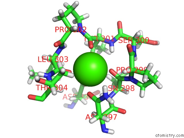
Mono view
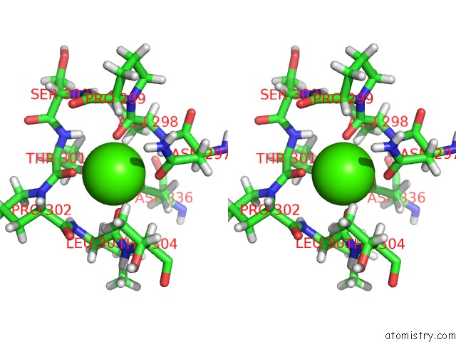
Stereo pair view
Mono view
Stereo pair view
A full contact list of Calcium with other atoms in the Ca binding
site number 1 of Crystal Structure of Three N-Terminal Domains of the Type V Pili Tip Protein MFA5 From Porphyromonas Gingivalis within 5.0Å range:
|
Reference:
T.V.Heidler,
R.Claesson,
K.Persson.
Structure of Porphyromonas Gingivalis MFA5 Reveals Unexpected Divergence For A Gram-Negative Fimbrial Tip Protein To Be Published.
Page generated: Tue Jul 16 15:23:54 2024
Last articles
Zn in 9J0NZn in 9J0O
Zn in 9J0P
Zn in 9FJX
Zn in 9EKB
Zn in 9C0F
Zn in 9CAH
Zn in 9CH0
Zn in 9CH3
Zn in 9CH1