Calcium »
PDB 6toy-6tz7 »
6tvr »
Calcium in PDB 6tvr: Crystal Structure of the Haemagglutinin Mutant (GLN226LEU) From An H10N7 Seal Influenza Virus Isolated in Germany
Protein crystallography data
The structure of Crystal Structure of the Haemagglutinin Mutant (GLN226LEU) From An H10N7 Seal Influenza Virus Isolated in Germany, PDB code: 6tvr
was solved by
J.Zhang,
X.Xiong,
A.Purkiss,
P.Walker,
S.Gamblin,
J.J.Skehel,
with X-Ray Crystallography technique. A brief refinement statistics is given in the table below:
| Resolution Low / High (Å) | 72.33 / 2.63 |
| Space group | P 1 21 1 |
| Cell size a, b, c (Å), α, β, γ (°) | 69.354, 214.102, 157.165, 90.00, 102.06, 90.00 |
| R / Rfree (%) | 24.1 / 26.4 |
Calcium Binding Sites:
The binding sites of Calcium atom in the Crystal Structure of the Haemagglutinin Mutant (GLN226LEU) From An H10N7 Seal Influenza Virus Isolated in Germany
(pdb code 6tvr). This binding sites where shown within
5.0 Angstroms radius around Calcium atom.
In total 4 binding sites of Calcium where determined in the Crystal Structure of the Haemagglutinin Mutant (GLN226LEU) From An H10N7 Seal Influenza Virus Isolated in Germany, PDB code: 6tvr:
Jump to Calcium binding site number: 1; 2; 3; 4;
In total 4 binding sites of Calcium where determined in the Crystal Structure of the Haemagglutinin Mutant (GLN226LEU) From An H10N7 Seal Influenza Virus Isolated in Germany, PDB code: 6tvr:
Jump to Calcium binding site number: 1; 2; 3; 4;
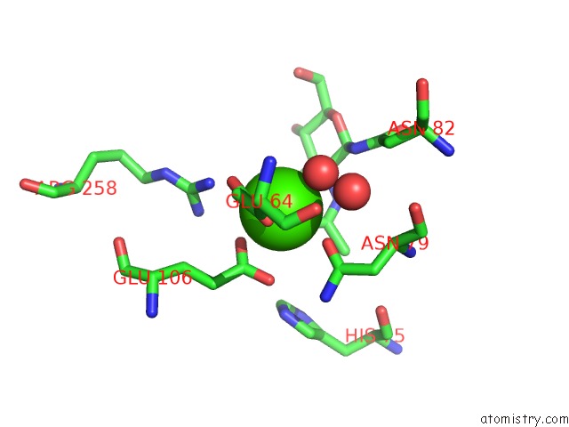
Calcium binding site 1 out of 4 in 6tvr
Go back to
Calcium binding site 1 out
of 4 in the Crystal Structure of the Haemagglutinin Mutant (GLN226LEU) From An H10N7 Seal Influenza Virus Isolated in Germany
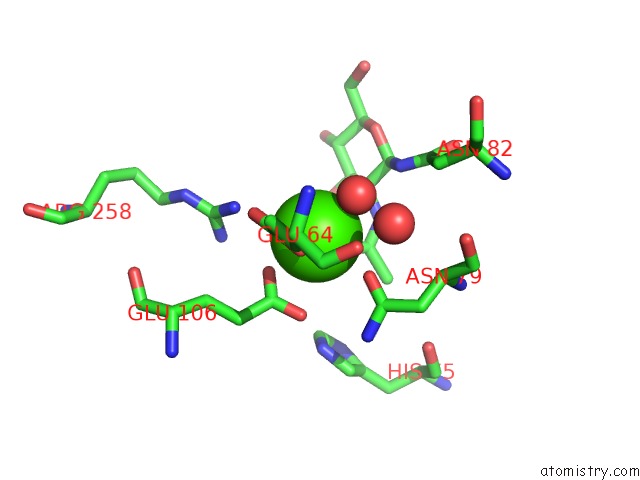
Mono view
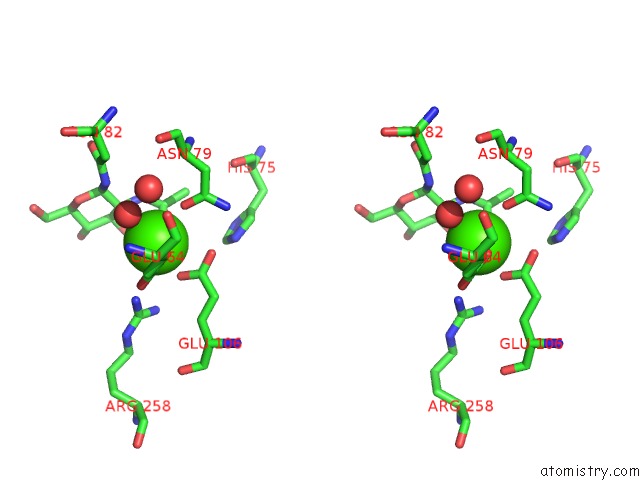
Stereo pair view
Mono view
Stereo pair view
A full contact list of Calcium with other atoms in the Ca binding
site number 1 of Crystal Structure of the Haemagglutinin Mutant (GLN226LEU) From An H10N7 Seal Influenza Virus Isolated in Germany within 5.0Å range:
|
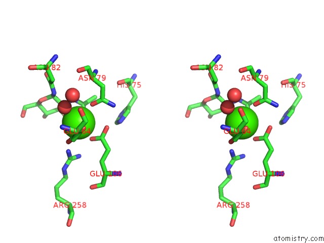
Calcium binding site 2 out of 4 in 6tvr
Go back to
Calcium binding site 2 out
of 4 in the Crystal Structure of the Haemagglutinin Mutant (GLN226LEU) From An H10N7 Seal Influenza Virus Isolated in Germany
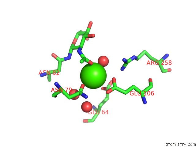
Mono view
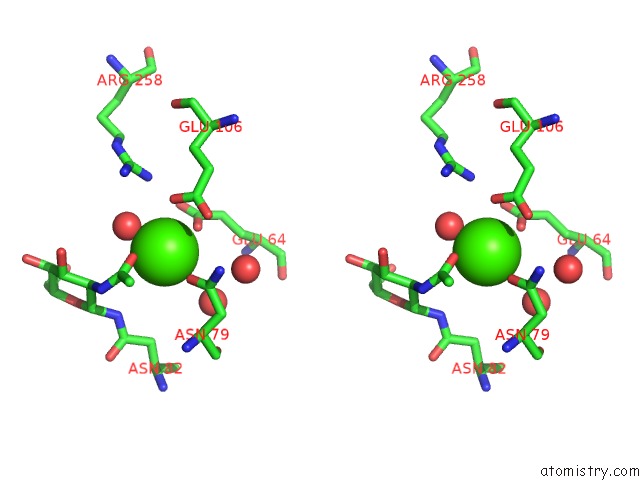
Stereo pair view
Mono view
Stereo pair view
A full contact list of Calcium with other atoms in the Ca binding
site number 2 of Crystal Structure of the Haemagglutinin Mutant (GLN226LEU) From An H10N7 Seal Influenza Virus Isolated in Germany within 5.0Å range:
|
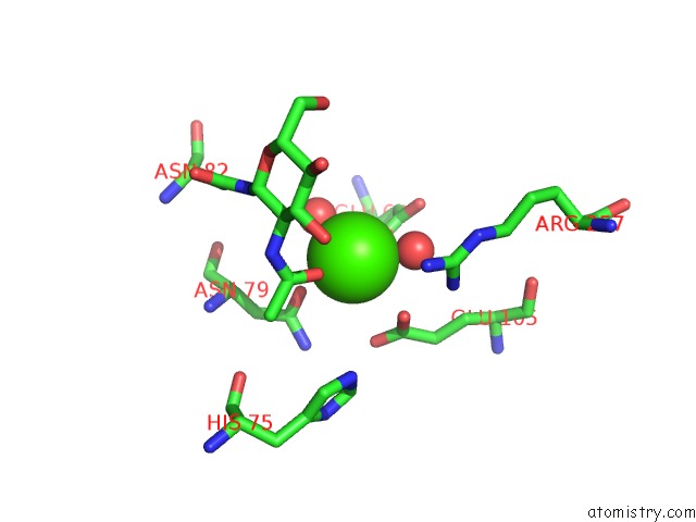
Calcium binding site 3 out of 4 in 6tvr
Go back to
Calcium binding site 3 out
of 4 in the Crystal Structure of the Haemagglutinin Mutant (GLN226LEU) From An H10N7 Seal Influenza Virus Isolated in Germany
Mono view
Stereo pair view
Mono view
Stereo pair view
A full contact list of Calcium with other atoms in the Ca binding
site number 3 of Crystal Structure of the Haemagglutinin Mutant (GLN226LEU) From An H10N7 Seal Influenza Virus Isolated in Germany within 5.0Å range:
|
Calcium binding site 4 out of 4 in 6tvr
Go back to
Calcium binding site 4 out
of 4 in the Crystal Structure of the Haemagglutinin Mutant (GLN226LEU) From An H10N7 Seal Influenza Virus Isolated in Germany
Mono view
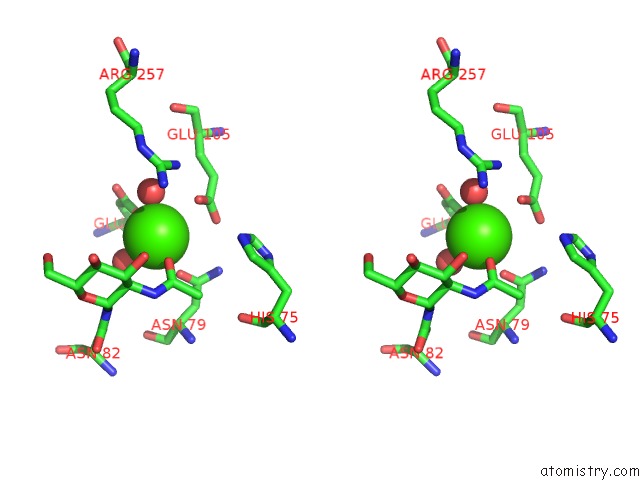
Stereo pair view
Mono view
Stereo pair view
A full contact list of Calcium with other atoms in the Ca binding
site number 4 of Crystal Structure of the Haemagglutinin Mutant (GLN226LEU) From An H10N7 Seal Influenza Virus Isolated in Germany within 5.0Å range:
|
Reference:
S.Herfst,
J.Zhang,
M.Richard,
R.Mcbride,
P.Lexmond,
T.M.Bestebroer,
M.I.J.Spronken,
D.De Meulder,
J.M.Van Den Brand,
M.E.Rosu,
S.R.Martin,
S.J.Gamblin,
X.Xiong,
W.Peng,
R.Bodewes,
E.Van Der Vries,
A.D.M.E.Osterhaus,
J.C.Paulson,
J.J.Skehel,
R.A.M.Fouchier.
Hemagglutinin Traits Determine Transmission of Avian A/H10N7 Influenza Virus Between Mammals. Cell Host Microbe V. 28 602 2020.
ISSN: ESSN 1934-6069
PubMed: 33031770
DOI: 10.1016/J.CHOM.2020.08.011
Page generated: Tue Jul 16 15:30:49 2024
ISSN: ESSN 1934-6069
PubMed: 33031770
DOI: 10.1016/J.CHOM.2020.08.011
Last articles
Zn in 9MJ5Zn in 9HNW
Zn in 9G0L
Zn in 9FNE
Zn in 9DZN
Zn in 9E0I
Zn in 9D32
Zn in 9DAK
Zn in 8ZXC
Zn in 8ZUF