Calcium »
PDB 6ulm-6v4o »
6uw1 »
Calcium in PDB 6uw1: The Crystal Structure of Fbia From Mycobacterium Smegmatis, Fo Bound Form
Enzymatic activity of The Crystal Structure of Fbia From Mycobacterium Smegmatis, Fo Bound Form
All present enzymatic activity of The Crystal Structure of Fbia From Mycobacterium Smegmatis, Fo Bound Form:
2.7.8.28;
2.7.8.28;
Protein crystallography data
The structure of The Crystal Structure of Fbia From Mycobacterium Smegmatis, Fo Bound Form, PDB code: 6uw1
was solved by
R.Grinter,
D.Gillet,
P.F.Cordero,
C.Greening,
with X-Ray Crystallography technique. A brief refinement statistics is given in the table below:
| Resolution Low / High (Å) | 45.90 / 2.21 |
| Space group | P 1 21 1 |
| Cell size a, b, c (Å), α, β, γ (°) | 46.065, 73.843, 91.699, 90.00, 94.91, 90.00 |
| R / Rfree (%) | 18.4 / 24.5 |
Calcium Binding Sites:
The binding sites of Calcium atom in the The Crystal Structure of Fbia From Mycobacterium Smegmatis, Fo Bound Form
(pdb code 6uw1). This binding sites where shown within
5.0 Angstroms radius around Calcium atom.
In total 2 binding sites of Calcium where determined in the The Crystal Structure of Fbia From Mycobacterium Smegmatis, Fo Bound Form, PDB code: 6uw1:
Jump to Calcium binding site number: 1; 2;
In total 2 binding sites of Calcium where determined in the The Crystal Structure of Fbia From Mycobacterium Smegmatis, Fo Bound Form, PDB code: 6uw1:
Jump to Calcium binding site number: 1; 2;
Calcium binding site 1 out of 2 in 6uw1
Go back to
Calcium binding site 1 out
of 2 in the The Crystal Structure of Fbia From Mycobacterium Smegmatis, Fo Bound Form
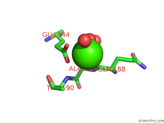
Mono view
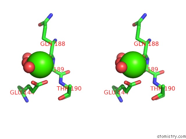
Stereo pair view
Mono view
Stereo pair view
A full contact list of Calcium with other atoms in the Ca binding
site number 1 of The Crystal Structure of Fbia From Mycobacterium Smegmatis, Fo Bound Form within 5.0Å range:
|
Calcium binding site 2 out of 2 in 6uw1
Go back to
Calcium binding site 2 out
of 2 in the The Crystal Structure of Fbia From Mycobacterium Smegmatis, Fo Bound Form
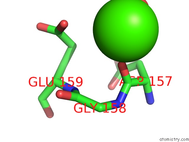
Mono view
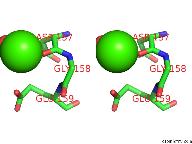
Stereo pair view
Mono view
Stereo pair view
A full contact list of Calcium with other atoms in the Ca binding
site number 2 of The Crystal Structure of Fbia From Mycobacterium Smegmatis, Fo Bound Form within 5.0Å range:
|
Reference:
R.Grinter,
R.Ney,
R.Brammananth,
C.Barlow,
C.R.F.Cordero,
D.Gillet,
T.Izore,
P.Crellin,
M.Cryle,
R.Schittenhelm,
R.Coppel,
C.Greening.
Cellular and Structural Basis of Synthesis of the Unique Intermediate Dehydro-F420-0 in Mycobacteria Msystems 2020.
ISSN: ISSN 2379-5077
Page generated: Tue Jul 16 16:04:11 2024
ISSN: ISSN 2379-5077
Last articles
Zn in 9MJ5Zn in 9HNW
Zn in 9G0L
Zn in 9FNE
Zn in 9DZN
Zn in 9E0I
Zn in 9D32
Zn in 9DAK
Zn in 8ZXC
Zn in 8ZUF