Calcium »
PDB 6ulw-6v4p »
6v2m »
Calcium in PDB 6v2m: Structure of Escherichia Coli ASP269ASN Mutant Phosphoenolpyruvate Carboxykinase
Enzymatic activity of Structure of Escherichia Coli ASP269ASN Mutant Phosphoenolpyruvate Carboxykinase
All present enzymatic activity of Structure of Escherichia Coli ASP269ASN Mutant Phosphoenolpyruvate Carboxykinase:
4.1.1.49;
4.1.1.49;
Protein crystallography data
The structure of Structure of Escherichia Coli ASP269ASN Mutant Phosphoenolpyruvate Carboxykinase, PDB code: 6v2m
was solved by
A.S.Sokaribo,
J.H.Cotelesage,
with X-Ray Crystallography technique. A brief refinement statistics is given in the table below:
| Resolution Low / High (Å) | 46.38 / 1.66 |
| Space group | C 1 2 1 |
| Cell size a, b, c (Å), α, β, γ (°) | 124.780, 94.220, 46.540, 90.00, 94.79, 90.00 |
| R / Rfree (%) | 18.5 / n/a |
Other elements in 6v2m:
The structure of Structure of Escherichia Coli ASP269ASN Mutant Phosphoenolpyruvate Carboxykinase also contains other interesting chemical elements:
| Magnesium | (Mg) | 1 atom |
Calcium Binding Sites:
The binding sites of Calcium atom in the Structure of Escherichia Coli ASP269ASN Mutant Phosphoenolpyruvate Carboxykinase
(pdb code 6v2m). This binding sites where shown within
5.0 Angstroms radius around Calcium atom.
In total only one binding site of Calcium was determined in the Structure of Escherichia Coli ASP269ASN Mutant Phosphoenolpyruvate Carboxykinase, PDB code: 6v2m:
In total only one binding site of Calcium was determined in the Structure of Escherichia Coli ASP269ASN Mutant Phosphoenolpyruvate Carboxykinase, PDB code: 6v2m:
Calcium binding site 1 out of 1 in 6v2m
Go back to
Calcium binding site 1 out
of 1 in the Structure of Escherichia Coli ASP269ASN Mutant Phosphoenolpyruvate Carboxykinase
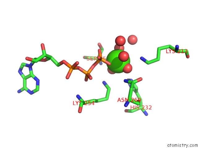
Mono view
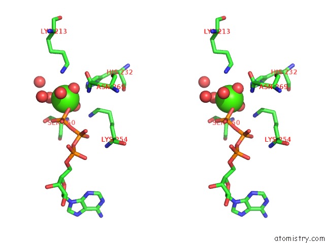
Stereo pair view
Mono view
Stereo pair view
A full contact list of Calcium with other atoms in the Ca binding
site number 1 of Structure of Escherichia Coli ASP269ASN Mutant Phosphoenolpyruvate Carboxykinase within 5.0Å range:
|
Reference:
A.S.Sokaribo,
J.H.Cotelesage,
B.Novakovski,
H.Goldie,
D.Sanders.
Structure of Escherichia Coli ASP269ASN Mutant Phosphoenolpyruvate Carboxykinase To Be Published.
Page generated: Wed Jul 9 18:56:06 2025
Last articles
F in 4FDOF in 4FDN
F in 4FC0
F in 4FAT
F in 4F9Y
F in 4FA2
F in 4F9W
F in 4FAD
F in 4ESV
F in 4F5R