Calcium »
PDB 6v4o-6vg4 »
6vel »
Calcium in PDB 6vel: Crystal Structure of Human E-Cadherin Bound By Mouse Monoclonal Antibody 66E8FAB
Protein crystallography data
The structure of Crystal Structure of Human E-Cadherin Bound By Mouse Monoclonal Antibody 66E8FAB, PDB code: 6vel
was solved by
Seattle Structural Genomics Center For Infectious Disease,
Seattlestructural Genomics Center For Infectious Disease (Ssgcid),
with X-Ray Crystallography technique. A brief refinement statistics is given in the table below:
| Resolution Low / High (Å) | 46.54 / 2.65 |
| Space group | P 31 2 1 |
| Cell size a, b, c (Å), α, β, γ (°) | 142.170, 142.170, 90.320, 90.00, 90.00, 120.00 |
| R / Rfree (%) | 18.3 / 23.3 |
Other elements in 6vel:
The structure of Crystal Structure of Human E-Cadherin Bound By Mouse Monoclonal Antibody 66E8FAB also contains other interesting chemical elements:
| Sodium | (Na) | 1 atom |
Calcium Binding Sites:
The binding sites of Calcium atom in the Crystal Structure of Human E-Cadherin Bound By Mouse Monoclonal Antibody 66E8FAB
(pdb code 6vel). This binding sites where shown within
5.0 Angstroms radius around Calcium atom.
In total 2 binding sites of Calcium where determined in the Crystal Structure of Human E-Cadherin Bound By Mouse Monoclonal Antibody 66E8FAB, PDB code: 6vel:
Jump to Calcium binding site number: 1; 2;
In total 2 binding sites of Calcium where determined in the Crystal Structure of Human E-Cadherin Bound By Mouse Monoclonal Antibody 66E8FAB, PDB code: 6vel:
Jump to Calcium binding site number: 1; 2;
Calcium binding site 1 out of 2 in 6vel
Go back to
Calcium binding site 1 out
of 2 in the Crystal Structure of Human E-Cadherin Bound By Mouse Monoclonal Antibody 66E8FAB

Mono view
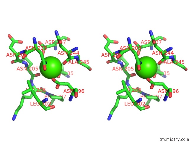
Stereo pair view
Mono view
Stereo pair view
A full contact list of Calcium with other atoms in the Ca binding
site number 1 of Crystal Structure of Human E-Cadherin Bound By Mouse Monoclonal Antibody 66E8FAB within 5.0Å range:
|
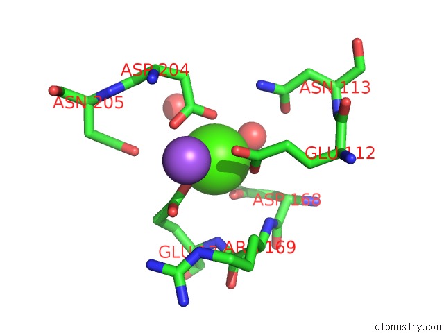
Calcium binding site 2 out of 2 in 6vel
Go back to
Calcium binding site 2 out
of 2 in the Crystal Structure of Human E-Cadherin Bound By Mouse Monoclonal Antibody 66E8FAB
Mono view
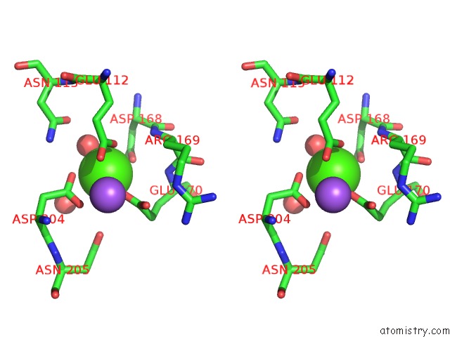
Stereo pair view
Mono view
Stereo pair view
A full contact list of Calcium with other atoms in the Ca binding
site number 2 of Crystal Structure of Human E-Cadherin Bound By Mouse Monoclonal Antibody 66E8FAB within 5.0Å range:
|
Reference:
M.J.Bolejack,
J.Abendroth,
A.Y.Maker,
B.M.Gumbiner,
D.D.Lorimer,
P.S.Horanyi,
T.E.Edwards.
Crystal Structure of Human E-Cadherin Bound By Mouse Monoclonal Antibody 66E8FAB To Be Published.
Page generated: Tue Jul 16 16:40:42 2024
Last articles
Zn in 9JYWZn in 9IR4
Zn in 9IR3
Zn in 9GMX
Zn in 9GMW
Zn in 9JEJ
Zn in 9ERF
Zn in 9ERE
Zn in 9EGV
Zn in 9EGW