Calcium »
PDB 6wni-6x6q »
6wnu »
Calcium in PDB 6wnu: Crystal Structure of the Three-Domain Cyclomaltodextrin Glucanotransferase Clda in the Monomeric Form
Enzymatic activity of Crystal Structure of the Three-Domain Cyclomaltodextrin Glucanotransferase Clda in the Monomeric Form
All present enzymatic activity of Crystal Structure of the Three-Domain Cyclomaltodextrin Glucanotransferase Clda in the Monomeric Form:
2.4.1.19;
2.4.1.19;
Protein crystallography data
The structure of Crystal Structure of the Three-Domain Cyclomaltodextrin Glucanotransferase Clda in the Monomeric Form, PDB code: 6wnu
was solved by
E.Magana-Cuevas,
S.Centeno-Leija,
H.Serrano-Posada,
with X-Ray Crystallography technique. A brief refinement statistics is given in the table below:
| Resolution Low / High (Å) | 30.08 / 1.88 |
| Space group | P 31 |
| Cell size a, b, c (Å), α, β, γ (°) | 67.77, 67.77, 105.1, 90, 90, 120 |
| R / Rfree (%) | 18.6 / 22.7 |
Calcium Binding Sites:
The binding sites of Calcium atom in the Crystal Structure of the Three-Domain Cyclomaltodextrin Glucanotransferase Clda in the Monomeric Form
(pdb code 6wnu). This binding sites where shown within
5.0 Angstroms radius around Calcium atom.
In total 3 binding sites of Calcium where determined in the Crystal Structure of the Three-Domain Cyclomaltodextrin Glucanotransferase Clda in the Monomeric Form, PDB code: 6wnu:
Jump to Calcium binding site number: 1; 2; 3;
In total 3 binding sites of Calcium where determined in the Crystal Structure of the Three-Domain Cyclomaltodextrin Glucanotransferase Clda in the Monomeric Form, PDB code: 6wnu:
Jump to Calcium binding site number: 1; 2; 3;
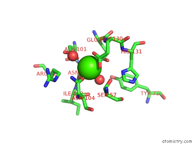
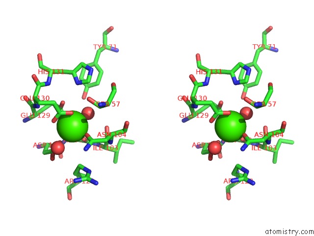
Calcium binding site 1 out of 3 in 6wnu
Go back to
Calcium binding site 1 out
of 3 in the Crystal Structure of the Three-Domain Cyclomaltodextrin Glucanotransferase Clda in the Monomeric Form
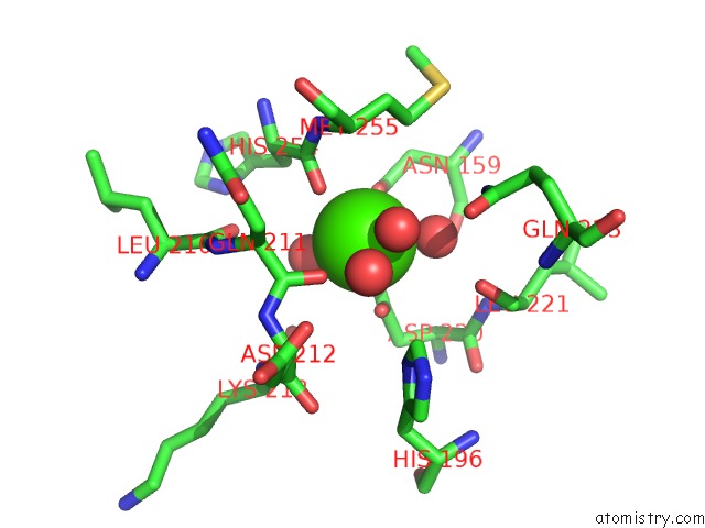
Mono view
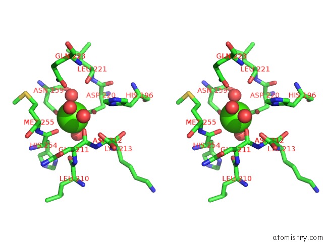
Stereo pair view
Mono view
Stereo pair view
A full contact list of Calcium with other atoms in the Ca binding
site number 1 of Crystal Structure of the Three-Domain Cyclomaltodextrin Glucanotransferase Clda in the Monomeric Form within 5.0Å range:
|
Calcium binding site 2 out of 3 in 6wnu
Go back to
Calcium binding site 2 out
of 3 in the Crystal Structure of the Three-Domain Cyclomaltodextrin Glucanotransferase Clda in the Monomeric Form
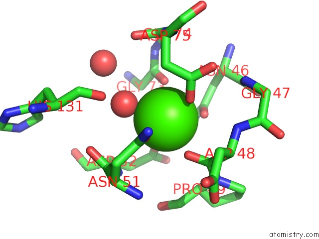
Mono view
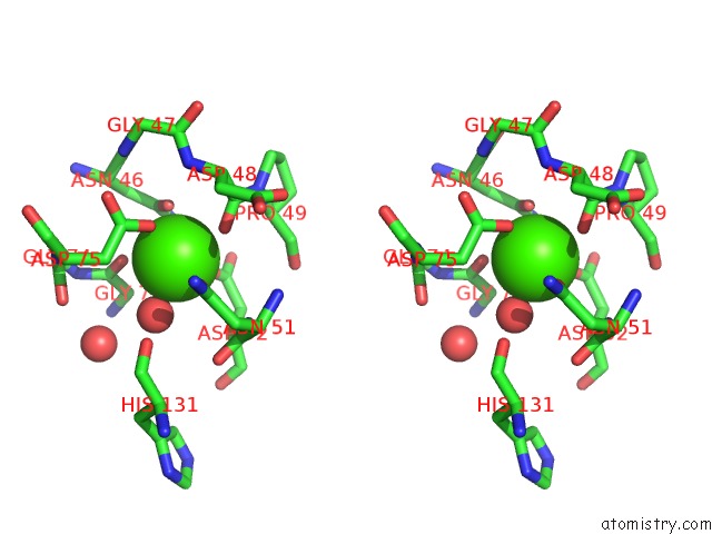
Stereo pair view
Mono view
Stereo pair view
A full contact list of Calcium with other atoms in the Ca binding
site number 2 of Crystal Structure of the Three-Domain Cyclomaltodextrin Glucanotransferase Clda in the Monomeric Form within 5.0Å range:
|
Calcium binding site 3 out of 3 in 6wnu
Go back to
Calcium binding site 3 out
of 3 in the Crystal Structure of the Three-Domain Cyclomaltodextrin Glucanotransferase Clda in the Monomeric Form
Mono view
Stereo pair view
Mono view
Stereo pair view
A full contact list of Calcium with other atoms in the Ca binding
site number 3 of Crystal Structure of the Three-Domain Cyclomaltodextrin Glucanotransferase Clda in the Monomeric Form within 5.0Å range:
|
Reference:
S.Centeno-Leija,
A.Lopez-Munguia,
Y.Cardenas-Conejo,
N.A.Mancilla-Margalli,
B.Velazquez-Cruz,
E.Magana-Cuevas,
Y.Guerra-Borrego,
R.Zatarain-Palacios,
Y.Marin-Tovar,
S.Gomez-Manzo,
J.Marcial-Quino,
B.Hernandez-Ochoa,
J.A.Osuna-Castro,
E.Rudino-Pinera,
H.Serrano-Posada.
Discovery of A Novel Group of Three-Domain Thermophilic Cyclomaltodextrin Glucanotransferases: Structural and Functional Implications. To Be Published.
Page generated: Wed Jul 9 19:37:37 2025
Last articles
Ca in 7M22Ca in 7LYU
Ca in 7LZ8
Ca in 7M0A
Ca in 7LZ7
Ca in 7M09
Ca in 7LX0
Ca in 7LVV
Ca in 7LXJ
Ca in 7LXH