Calcium »
PDB 6wni-6x6q »
6wrv »
Calcium in PDB 6wrv: Crystal Structure of Computationally Designed Protein 3DS18 in Complex with the Human Transferrin Receptor Ectodomain
Protein crystallography data
The structure of Crystal Structure of Computationally Designed Protein 3DS18 in Complex with the Human Transferrin Receptor Ectodomain, PDB code: 6wrv
was solved by
J.Abraham,
D.Baker,
D.D.Sahtoe,
A.Coscia,
L.Clark,
D.Olal,
with X-Ray Crystallography technique. A brief refinement statistics is given in the table below:
| Resolution Low / High (Å) | 128.04 / 2.47 |
| Space group | P 42 21 2 |
| Cell size a, b, c (Å), α, β, γ (°) | 256.079, 256.079, 128.084, 90, 90, 90 |
| R / Rfree (%) | 22.1 / 23.6 |
Calcium Binding Sites:
The binding sites of Calcium atom in the Crystal Structure of Computationally Designed Protein 3DS18 in Complex with the Human Transferrin Receptor Ectodomain
(pdb code 6wrv). This binding sites where shown within
5.0 Angstroms radius around Calcium atom.
In total 3 binding sites of Calcium where determined in the Crystal Structure of Computationally Designed Protein 3DS18 in Complex with the Human Transferrin Receptor Ectodomain, PDB code: 6wrv:
Jump to Calcium binding site number: 1; 2; 3;
In total 3 binding sites of Calcium where determined in the Crystal Structure of Computationally Designed Protein 3DS18 in Complex with the Human Transferrin Receptor Ectodomain, PDB code: 6wrv:
Jump to Calcium binding site number: 1; 2; 3;
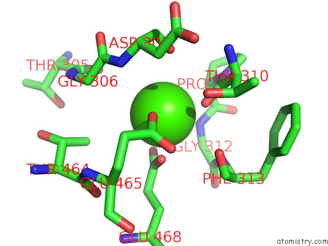
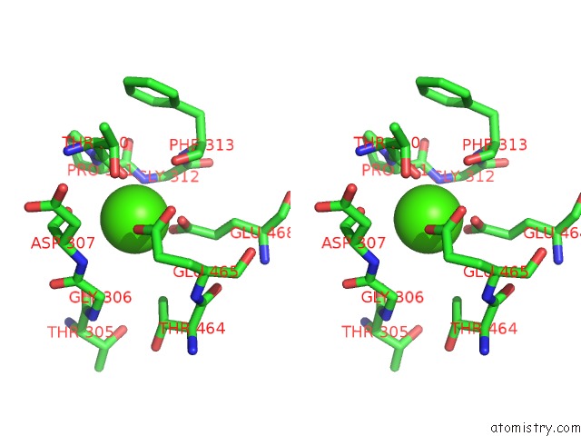
Calcium binding site 1 out of 3 in 6wrv
Go back to
Calcium binding site 1 out
of 3 in the Crystal Structure of Computationally Designed Protein 3DS18 in Complex with the Human Transferrin Receptor Ectodomain
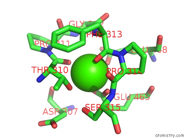
Mono view
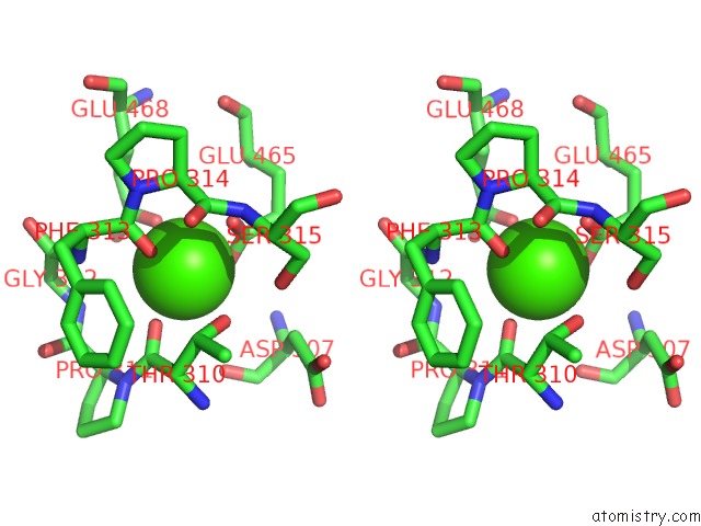
Stereo pair view
Mono view
Stereo pair view
A full contact list of Calcium with other atoms in the Ca binding
site number 1 of Crystal Structure of Computationally Designed Protein 3DS18 in Complex with the Human Transferrin Receptor Ectodomain within 5.0Å range:
|
Calcium binding site 2 out of 3 in 6wrv
Go back to
Calcium binding site 2 out
of 3 in the Crystal Structure of Computationally Designed Protein 3DS18 in Complex with the Human Transferrin Receptor Ectodomain
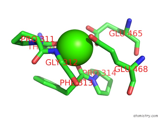
Mono view
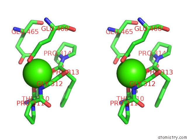
Stereo pair view
Mono view
Stereo pair view
A full contact list of Calcium with other atoms in the Ca binding
site number 2 of Crystal Structure of Computationally Designed Protein 3DS18 in Complex with the Human Transferrin Receptor Ectodomain within 5.0Å range:
|
Calcium binding site 3 out of 3 in 6wrv
Go back to
Calcium binding site 3 out
of 3 in the Crystal Structure of Computationally Designed Protein 3DS18 in Complex with the Human Transferrin Receptor Ectodomain
Mono view
Stereo pair view
Mono view
Stereo pair view
A full contact list of Calcium with other atoms in the Ca binding
site number 3 of Crystal Structure of Computationally Designed Protein 3DS18 in Complex with the Human Transferrin Receptor Ectodomain within 5.0Å range:
|
Reference:
D.D.Sahtoe,
A.Coscia,
N.Mustafaoglu,
L.M.Miller,
D.Olal,
I.Vulovic,
T.Y.Yu,
I.Goreshnik,
Y.R.Lin,
L.Clark,
F.Busch,
L.Stewart,
V.H.Wysocki,
D.E.Ingber,
J.Abraham,
D.Baker.
Transferrin Receptor Targeting By De Novo Sheet Extension. Proc.Natl.Acad.Sci.Usa V. 118 2021.
ISSN: ESSN 1091-6490
PubMed: 33879614
DOI: 10.1073/PNAS.2021569118
Page generated: Wed Jul 9 19:37:52 2025
ISSN: ESSN 1091-6490
PubMed: 33879614
DOI: 10.1073/PNAS.2021569118
Last articles
Ca in 7NSDCa in 7NQC
Ca in 7NMI
Ca in 7NP8
Ca in 7NSA
Ca in 7NS9
Ca in 7NRK
Ca in 7NPG
Ca in 7NPB
Ca in 7NN9