Calcium in PDB 6x8a: Sucrose-Bound Structure of Marinomonas Primoryensis PA14 Carbohydrate- Binding Domain
Protein crystallography data
The structure of Sucrose-Bound Structure of Marinomonas Primoryensis PA14 Carbohydrate- Binding Domain, PDB code: 6x8a
was solved by
S.Guo,
P.L.Davies,
with X-Ray Crystallography technique. A brief refinement statistics is given in the table below:
Resolution Low / High (Å)
|
42.71 /
1.06
|
Space group
|
P 21 21 21
|
Cell size a, b, c (Å), α, β, γ (°)
|
45.23,
50.61,
79.6,
90,
90,
90
|
R / Rfree (%)
|
14.2 /
16.3
|
Calcium Binding Sites:
The binding sites of Calcium atom in the Sucrose-Bound Structure of Marinomonas Primoryensis PA14 Carbohydrate- Binding Domain
(pdb code 6x8a). This binding sites where shown within
5.0 Angstroms radius around Calcium atom.
In total 7 binding sites of Calcium where determined in the
Sucrose-Bound Structure of Marinomonas Primoryensis PA14 Carbohydrate- Binding Domain, PDB code: 6x8a:
Jump to Calcium binding site number:
1;
2;
3;
4;
5;
6;
7;
Calcium binding site 1 out
of 7 in 6x8a
Go back to
Calcium Binding Sites List in 6x8a
Calcium binding site 1 out
of 7 in the Sucrose-Bound Structure of Marinomonas Primoryensis PA14 Carbohydrate- Binding Domain
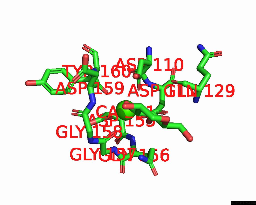
Mono view
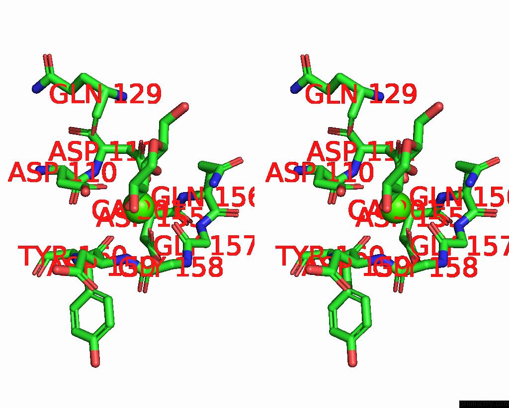
Stereo pair view
|
A full contact list of Calcium with other atoms in the Ca binding
site number 1 of Sucrose-Bound Structure of Marinomonas Primoryensis PA14 Carbohydrate- Binding Domain within 5.0Å range:
probe
|
atom
|
residue
|
distance (Å)
|
B
|
Occ
|
A:Ca201
b:4.4
occ:1.00
|
OD1
|
A:ASP110
|
2.3
|
5.0
|
1.0
|
OD1
|
A:ASP155
|
2.3
|
4.6
|
1.0
|
O
|
A:ASP159
|
2.4
|
5.0
|
1.0
|
O
|
A:GLY157
|
2.4
|
5.7
|
1.0
|
OD2
|
A:ASP111
|
2.4
|
4.5
|
1.0
|
O3
|
B:GLC1
|
2.5
|
6.3
|
1.0
|
O4
|
B:GLC1
|
2.5
|
6.1
|
1.0
|
CG
|
A:ASP110
|
3.3
|
5.1
|
1.0
|
CG
|
A:ASP111
|
3.3
|
4.6
|
1.0
|
C3
|
B:GLC1
|
3.3
|
8.0
|
1.0
|
C4
|
B:GLC1
|
3.4
|
7.6
|
1.0
|
C
|
A:GLY157
|
3.5
|
5.7
|
1.0
|
CG
|
A:ASP155
|
3.5
|
4.1
|
1.0
|
C
|
A:ASP159
|
3.5
|
5.3
|
1.0
|
OD2
|
A:ASP110
|
3.6
|
6.2
|
1.0
|
OD1
|
A:ASP111
|
3.7
|
5.1
|
1.0
|
N
|
A:ASP159
|
3.9
|
5.8
|
1.0
|
N
|
A:GLY157
|
4.0
|
5.2
|
1.0
|
C
|
A:GLY158
|
4.2
|
5.9
|
1.0
|
N
|
A:GLN156
|
4.3
|
4.2
|
1.0
|
CA
|
A:ASP159
|
4.3
|
5.9
|
1.0
|
N
|
A:GLY158
|
4.3
|
5.9
|
1.0
|
CA
|
A:GLY157
|
4.3
|
5.8
|
1.0
|
OD2
|
A:ASP155
|
4.3
|
5.2
|
1.0
|
CA
|
A:GLY158
|
4.4
|
6.0
|
1.0
|
CB
|
A:ASP155
|
4.4
|
4.3
|
1.0
|
CA
|
A:ASP155
|
4.4
|
3.9
|
1.0
|
CG
|
A:GLN156
|
4.5
|
6.9
|
1.0
|
N
|
A:ASP111
|
4.5
|
4.2
|
1.0
|
C
|
A:ASP110
|
4.5
|
3.9
|
1.0
|
NE2
|
A:GLN156
|
4.5
|
9.4
|
1.0
|
CB
|
A:ASP111
|
4.5
|
4.8
|
1.0
|
N
|
A:TYR160
|
4.6
|
5.2
|
1.0
|
CB
|
A:ASP110
|
4.6
|
5.0
|
1.0
|
O
|
A:ASP110
|
4.7
|
4.1
|
1.0
|
C2
|
B:GLC1
|
4.7
|
11.1
|
1.0
|
C5
|
B:GLC1
|
4.7
|
10.9
|
1.0
|
CA
|
A:TYR160
|
4.7
|
5.5
|
1.0
|
C
|
A:ASP155
|
4.8
|
4.1
|
1.0
|
O
|
A:GLY158
|
4.8
|
6.6
|
1.0
|
C
|
A:GLN156
|
4.9
|
5.0
|
1.0
|
CA
|
A:ASP111
|
4.9
|
4.4
|
1.0
|
O
|
A:GLN129
|
5.0
|
6.8
|
1.0
|
|
Calcium binding site 2 out
of 7 in 6x8a
Go back to
Calcium Binding Sites List in 6x8a
Calcium binding site 2 out
of 7 in the Sucrose-Bound Structure of Marinomonas Primoryensis PA14 Carbohydrate- Binding Domain
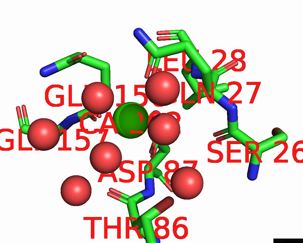
Mono view
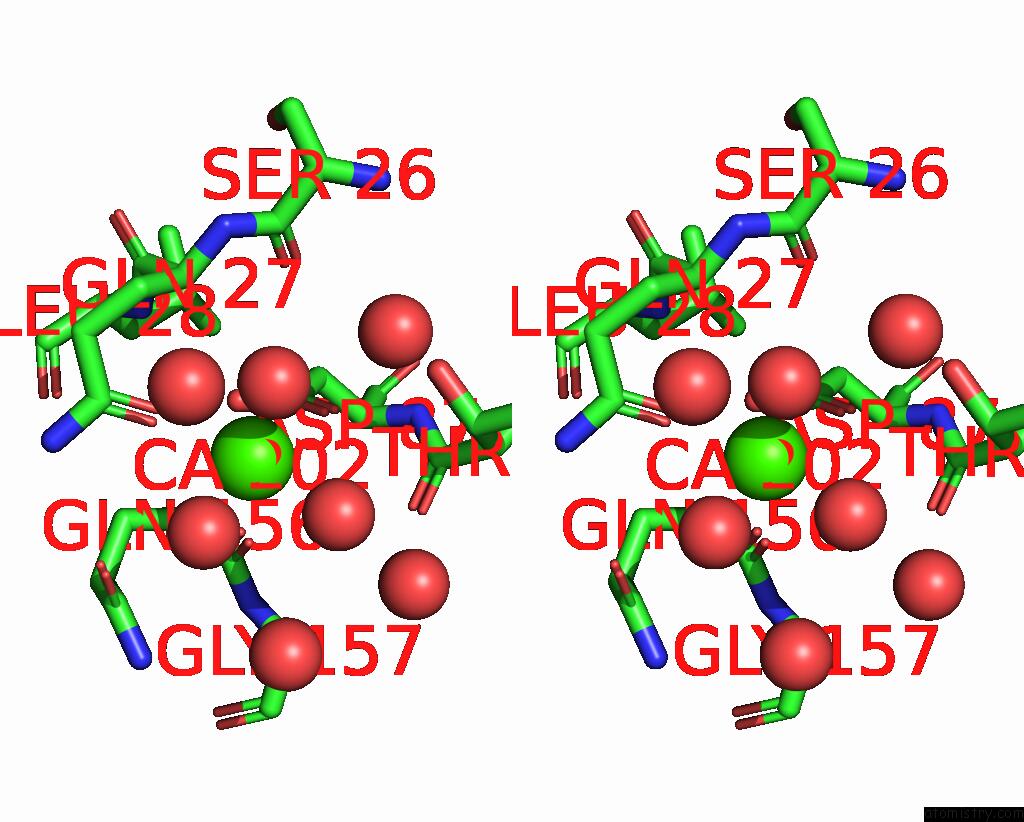
Stereo pair view
|
A full contact list of Calcium with other atoms in the Ca binding
site number 2 of Sucrose-Bound Structure of Marinomonas Primoryensis PA14 Carbohydrate- Binding Domain within 5.0Å range:
probe
|
atom
|
residue
|
distance (Å)
|
B
|
Occ
|
A:Ca202
b:6.5
occ:1.00
|
O
|
A:GLN156
|
2.3
|
6.3
|
1.0
|
OE1
|
A:GLN27
|
2.3
|
7.8
|
1.0
|
O
|
A:HOH479
|
2.4
|
8.6
|
1.0
|
O
|
A:HOH360
|
2.4
|
11.8
|
1.0
|
O
|
A:HOH497
|
2.4
|
9.4
|
1.0
|
OD1
|
A:ASP87
|
2.4
|
6.6
|
1.0
|
OD2
|
A:ASP87
|
2.5
|
5.8
|
1.0
|
CG
|
A:ASP87
|
2.8
|
5.6
|
1.0
|
C
|
A:GLN156
|
3.4
|
5.0
|
1.0
|
CD
|
A:GLN27
|
3.5
|
7.2
|
1.0
|
CA
|
A:GLN156
|
4.0
|
4.8
|
1.0
|
OE1
|
A:GLN156
|
4.0
|
10.2
|
1.0
|
CB
|
A:GLN156
|
4.2
|
5.7
|
1.0
|
CA
|
A:GLN27
|
4.2
|
6.2
|
1.0
|
NE2
|
A:GLN27
|
4.3
|
8.5
|
1.0
|
CB
|
A:ASP87
|
4.3
|
5.1
|
1.0
|
O
|
A:HOH516
|
4.4
|
11.9
|
1.0
|
O
|
A:HOH524
|
4.4
|
37.1
|
1.0
|
O
|
A:HOH380
|
4.4
|
9.0
|
1.0
|
CD
|
A:GLN156
|
4.5
|
8.6
|
1.0
|
CB
|
A:GLN27
|
4.5
|
7.0
|
1.0
|
O
|
A:HOH338
|
4.5
|
10.3
|
1.0
|
OG1
|
A:THR86
|
4.5
|
6.3
|
1.0
|
N
|
A:GLY157
|
4.6
|
5.2
|
1.0
|
CG
|
A:GLN27
|
4.6
|
7.5
|
1.0
|
O
|
A:SER26
|
4.7
|
8.2
|
1.0
|
O
|
A:THR86
|
4.8
|
6.0
|
1.0
|
N
|
A:LEU28
|
4.8
|
6.0
|
1.0
|
CA
|
A:GLY157
|
4.9
|
5.8
|
1.0
|
CG
|
A:GLN156
|
4.9
|
6.9
|
1.0
|
CB
|
A:THR86
|
5.0
|
6.4
|
1.0
|
|
Calcium binding site 3 out
of 7 in 6x8a
Go back to
Calcium Binding Sites List in 6x8a
Calcium binding site 3 out
of 7 in the Sucrose-Bound Structure of Marinomonas Primoryensis PA14 Carbohydrate- Binding Domain
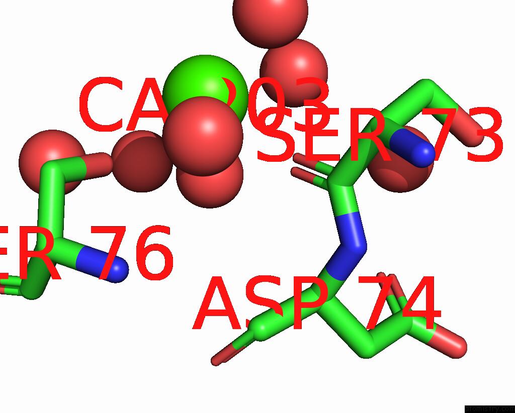
Mono view
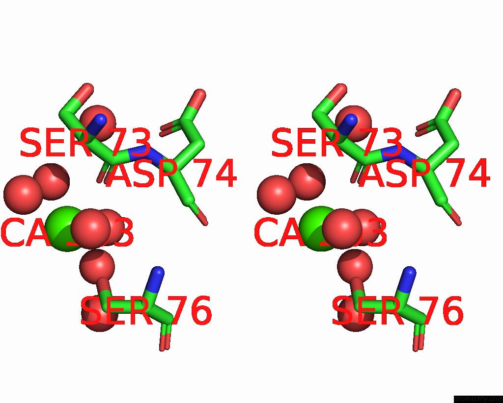
Stereo pair view
|
A full contact list of Calcium with other atoms in the Ca binding
site number 3 of Sucrose-Bound Structure of Marinomonas Primoryensis PA14 Carbohydrate- Binding Domain within 5.0Å range:
probe
|
atom
|
residue
|
distance (Å)
|
B
|
Occ
|
A:Ca203
b:10.4
occ:0.76
|
O
|
A:HOH496
|
2.3
|
20.2
|
1.0
|
O
|
A:SER73
|
2.4
|
11.0
|
1.0
|
O
|
A:HOH457
|
2.5
|
13.8
|
1.0
|
OG
|
A:SER76
|
2.6
|
8.6
|
1.0
|
O
|
A:HOH442
|
2.6
|
11.0
|
1.0
|
O
|
A:HOH418
|
2.6
|
21.1
|
1.0
|
C
|
A:SER73
|
3.4
|
9.6
|
1.0
|
CB
|
A:SER76
|
3.6
|
7.4
|
1.0
|
CA
|
A:SER73
|
4.0
|
10.3
|
1.0
|
O
|
A:HOH351
|
4.4
|
12.4
|
1.0
|
N
|
A:SER76
|
4.4
|
5.7
|
1.0
|
N
|
A:ASP74
|
4.5
|
7.3
|
1.0
|
CB
|
A:SER73
|
4.6
|
11.0
|
1.0
|
CA
|
A:SER76
|
4.6
|
6.2
|
1.0
|
O
|
A:HOH551
|
4.8
|
19.2
|
1.0
|
CA
|
A:ASP74
|
4.8
|
6.5
|
1.0
|
O
|
A:HOH341
|
4.9
|
14.9
|
1.0
|
C
|
A:ASP74
|
5.0
|
6.7
|
1.0
|
|
Calcium binding site 4 out
of 7 in 6x8a
Go back to
Calcium Binding Sites List in 6x8a
Calcium binding site 4 out
of 7 in the Sucrose-Bound Structure of Marinomonas Primoryensis PA14 Carbohydrate- Binding Domain
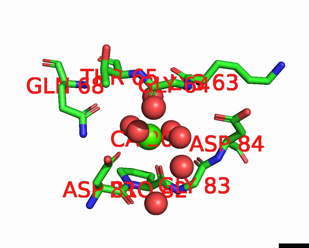
Mono view
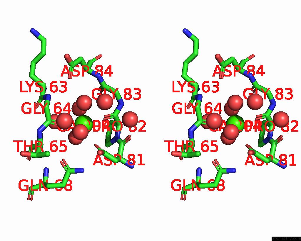
Stereo pair view
|
A full contact list of Calcium with other atoms in the Ca binding
site number 4 of Sucrose-Bound Structure of Marinomonas Primoryensis PA14 Carbohydrate- Binding Domain within 5.0Å range:
probe
|
atom
|
residue
|
distance (Å)
|
B
|
Occ
|
A:Ca204
b:5.6
occ:1.00
|
O
|
A:PRO82
|
2.3
|
6.2
|
1.0
|
O
|
A:HOH396
|
2.4
|
5.6
|
1.0
|
O
|
A:HOH336
|
2.4
|
11.9
|
1.0
|
O
|
A:GLY64
|
2.4
|
6.8
|
1.0
|
O
|
A:HOH408
|
2.4
|
8.2
|
1.0
|
OD1
|
A:ASP81
|
2.4
|
6.4
|
1.0
|
O
|
A:HOH414
|
2.4
|
7.5
|
1.0
|
CG
|
A:ASP81
|
3.5
|
6.1
|
1.0
|
C
|
A:GLY64
|
3.5
|
6.0
|
1.0
|
C
|
A:PRO82
|
3.5
|
6.4
|
1.0
|
OD2
|
A:ASP81
|
3.9
|
7.0
|
1.0
|
O
|
A:HOH327
|
4.1
|
5.4
|
1.0
|
N
|
A:PRO82
|
4.2
|
6.4
|
1.0
|
CA
|
A:GLY64
|
4.3
|
6.7
|
1.0
|
O
|
A:HOH539
|
4.3
|
37.3
|
1.0
|
CA
|
A:PRO82
|
4.4
|
6.9
|
1.0
|
C
|
A:ASP81
|
4.4
|
6.7
|
1.0
|
OE1
|
A:GLN68
|
4.4
|
6.0
|
1.0
|
N
|
A:GLY64
|
4.4
|
6.4
|
1.0
|
CD
|
A:PRO82
|
4.5
|
7.1
|
1.0
|
O
|
A:GLY83
|
4.5
|
6.4
|
1.0
|
N
|
A:GLY83
|
4.5
|
6.8
|
1.0
|
N
|
A:THR65
|
4.5
|
6.1
|
1.0
|
C
|
A:GLY83
|
4.5
|
5.9
|
1.0
|
CA
|
A:GLY83
|
4.6
|
7.0
|
1.0
|
NE2
|
A:GLN68
|
4.6
|
6.8
|
1.0
|
CA
|
A:THR65
|
4.7
|
6.2
|
1.0
|
O
|
A:ASP81
|
4.7
|
7.5
|
1.0
|
O
|
A:HOH535
|
4.7
|
16.6
|
1.0
|
CB
|
A:ASP81
|
4.8
|
6.4
|
1.0
|
CB
|
A:PRO82
|
4.8
|
9.7
|
1.0
|
OD1
|
A:ASP84
|
4.8
|
9.1
|
1.0
|
O
|
A:HOH420
|
4.9
|
31.9
|
1.0
|
CA
|
A:ASP81
|
4.9
|
6.6
|
1.0
|
C
|
A:LYS63
|
5.0
|
6.2
|
1.0
|
CD
|
A:GLN68
|
5.0
|
5.6
|
1.0
|
|
Calcium binding site 5 out
of 7 in 6x8a
Go back to
Calcium Binding Sites List in 6x8a
Calcium binding site 5 out
of 7 in the Sucrose-Bound Structure of Marinomonas Primoryensis PA14 Carbohydrate- Binding Domain
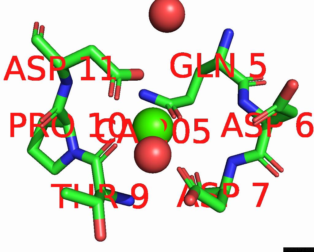
Mono view
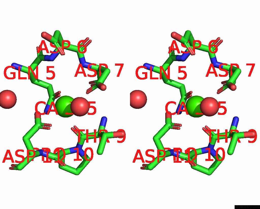
Stereo pair view
|
A full contact list of Calcium with other atoms in the Ca binding
site number 5 of Sucrose-Bound Structure of Marinomonas Primoryensis PA14 Carbohydrate- Binding Domain within 5.0Å range:
probe
|
atom
|
residue
|
distance (Å)
|
B
|
Occ
|
A:Ca205
b:21.9
occ:0.66
|
OD1
|
A:ASP7
|
2.3
|
38.8
|
1.0
|
O
|
A:HOH306
|
2.3
|
35.6
|
1.0
|
OE1
|
A:GLN5
|
2.4
|
24.5
|
1.0
|
O
|
A:THR9
|
2.4
|
20.8
|
1.0
|
OD2
|
A:ASP11
|
2.5
|
29.0
|
1.0
|
OD1
|
A:ASP11
|
2.5
|
28.2
|
1.0
|
CG
|
A:ASP11
|
2.8
|
22.8
|
1.0
|
CG
|
A:ASP7
|
3.3
|
37.0
|
1.0
|
CD
|
A:GLN5
|
3.3
|
23.6
|
1.0
|
C
|
A:THR9
|
3.6
|
21.1
|
1.0
|
NE2
|
A:GLN5
|
3.6
|
23.6
|
1.0
|
OD2
|
A:ASP7
|
3.7
|
44.2
|
1.0
|
CB
|
A:THR9
|
4.1
|
31.4
|
1.0
|
CA
|
A:THR9
|
4.2
|
26.0
|
1.0
|
N
|
A:THR9
|
4.2
|
26.7
|
1.0
|
CB
|
A:ASP11
|
4.3
|
18.8
|
1.0
|
N
|
A:ASP7
|
4.4
|
32.4
|
1.0
|
OD1
|
A:ASP6
|
4.5
|
56.2
|
1.0
|
N
|
A:ASP11
|
4.6
|
17.3
|
1.0
|
C
|
A:PRO10
|
4.6
|
18.0
|
1.0
|
CB
|
A:ASP7
|
4.6
|
30.9
|
1.0
|
N
|
A:PRO10
|
4.7
|
17.9
|
1.0
|
CG2
|
A:THR9
|
4.7
|
34.0
|
1.0
|
N
|
A:ASP6
|
4.7
|
33.2
|
1.0
|
CG
|
A:GLN5
|
4.7
|
26.8
|
1.0
|
CA
|
A:ASP7
|
4.8
|
30.5
|
1.0
|
C
|
A:ASP7
|
4.8
|
29.4
|
1.0
|
CA
|
A:ASP11
|
4.9
|
15.1
|
1.0
|
CA
|
A:PRO10
|
4.9
|
17.6
|
1.0
|
O
|
A:ASP7
|
4.9
|
30.6
|
1.0
|
O
|
A:PRO10
|
5.0
|
22.7
|
1.0
|
O
|
A:HOH490
|
5.0
|
31.4
|
1.0
|
|
Calcium binding site 6 out
of 7 in 6x8a
Go back to
Calcium Binding Sites List in 6x8a
Calcium binding site 6 out
of 7 in the Sucrose-Bound Structure of Marinomonas Primoryensis PA14 Carbohydrate- Binding Domain
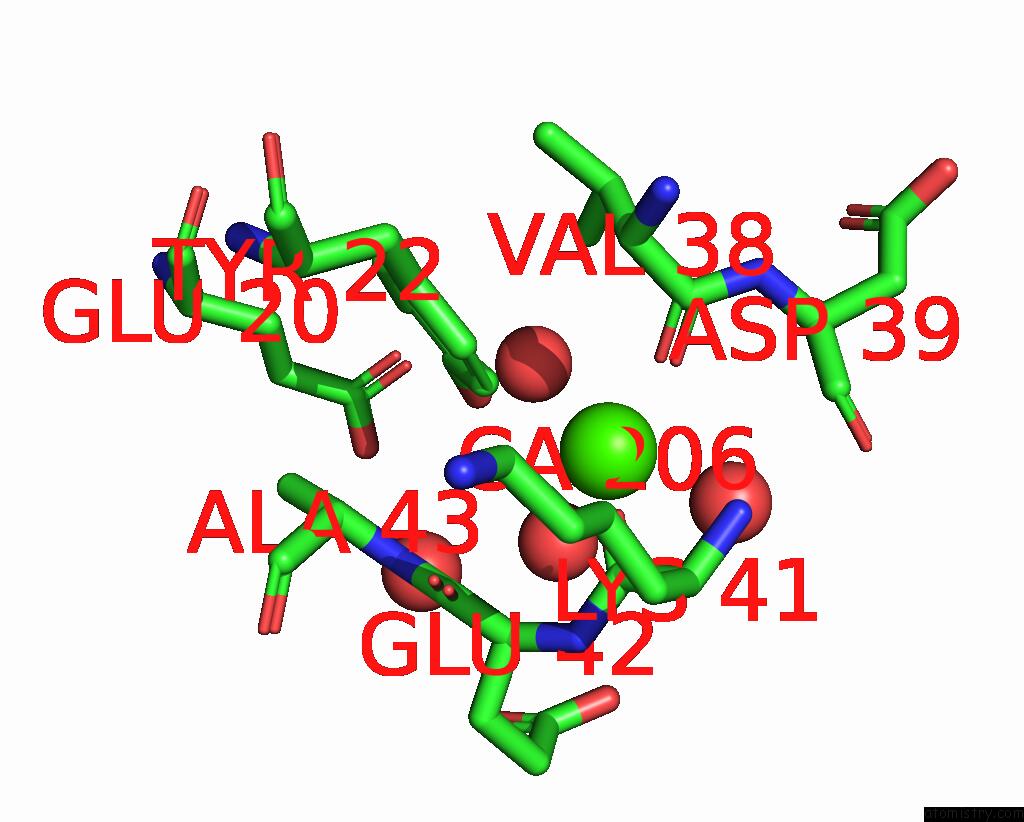
Mono view
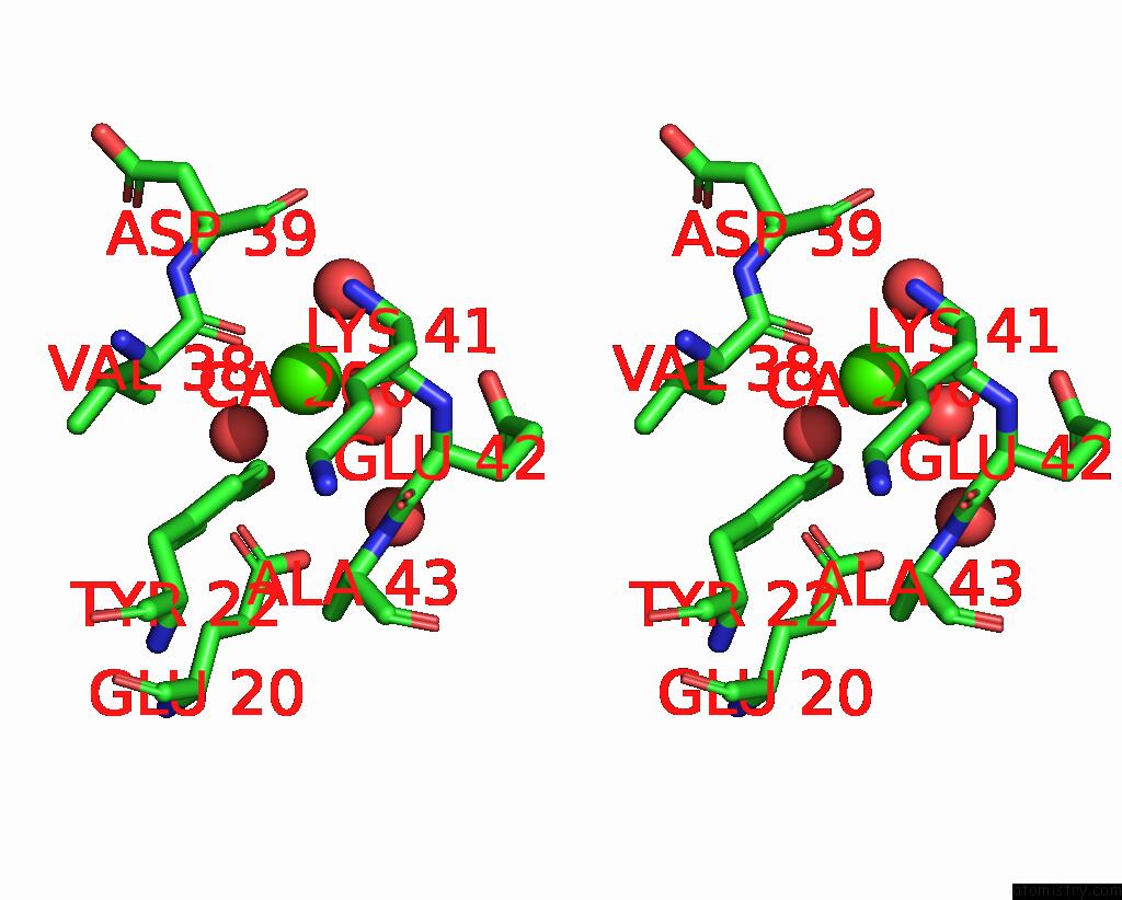
Stereo pair view
|
A full contact list of Calcium with other atoms in the Ca binding
site number 6 of Sucrose-Bound Structure of Marinomonas Primoryensis PA14 Carbohydrate- Binding Domain within 5.0Å range:
probe
|
atom
|
residue
|
distance (Å)
|
B
|
Occ
|
A:Ca206
b:22.1
occ:1.00
|
O
|
A:VAL38
|
2.4
|
10.3
|
1.0
|
O
|
A:HOH494
|
2.4
|
33.2
|
1.0
|
O
|
A:LYS41
|
2.4
|
9.8
|
1.0
|
O
|
A:HOH487
|
2.5
|
26.7
|
1.0
|
OH
|
A:TYR22
|
2.7
|
12.7
|
1.0
|
CZ
|
A:TYR22
|
3.5
|
9.4
|
1.0
|
C
|
A:VAL38
|
3.6
|
9.0
|
1.0
|
C
|
A:LYS41
|
3.6
|
9.9
|
1.0
|
CE2
|
A:TYR22
|
3.6
|
8.9
|
1.0
|
O
|
A:HOH448
|
3.9
|
26.4
|
1.0
|
CG1
|
A:VAL38
|
4.1
|
9.0
|
1.0
|
CA
|
A:GLU42
|
4.3
|
11.8
|
1.0
|
O
|
A:HOH429
|
4.4
|
21.1
|
1.0
|
N
|
A:GLU42
|
4.4
|
11.1
|
1.0
|
CA
|
A:VAL38
|
4.4
|
7.5
|
1.0
|
N
|
A:LYS41
|
4.4
|
10.4
|
1.0
|
N
|
A:ASP39
|
4.5
|
9.0
|
1.0
|
CA
|
A:ASP39
|
4.6
|
11.5
|
1.0
|
C
|
A:ASP39
|
4.6
|
10.9
|
1.0
|
N
|
A:ALA43
|
4.7
|
9.4
|
1.0
|
C
|
A:GLU42
|
4.7
|
10.7
|
1.0
|
CA
|
A:LYS41
|
4.7
|
10.8
|
1.0
|
OE2
|
A:GLU42
|
4.7
|
35.4
|
1.0
|
OE1
|
A:GLU20
|
4.7
|
13.2
|
1.0
|
CE1
|
A:TYR22
|
4.8
|
7.3
|
1.0
|
O
|
A:ASP39
|
4.9
|
14.3
|
1.0
|
CD2
|
A:TYR22
|
4.9
|
7.7
|
1.0
|
CB
|
A:VAL38
|
5.0
|
8.4
|
1.0
|
|
Calcium binding site 7 out
of 7 in 6x8a
Go back to
Calcium Binding Sites List in 6x8a
Calcium binding site 7 out
of 7 in the Sucrose-Bound Structure of Marinomonas Primoryensis PA14 Carbohydrate- Binding Domain
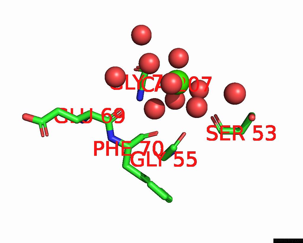
Mono view
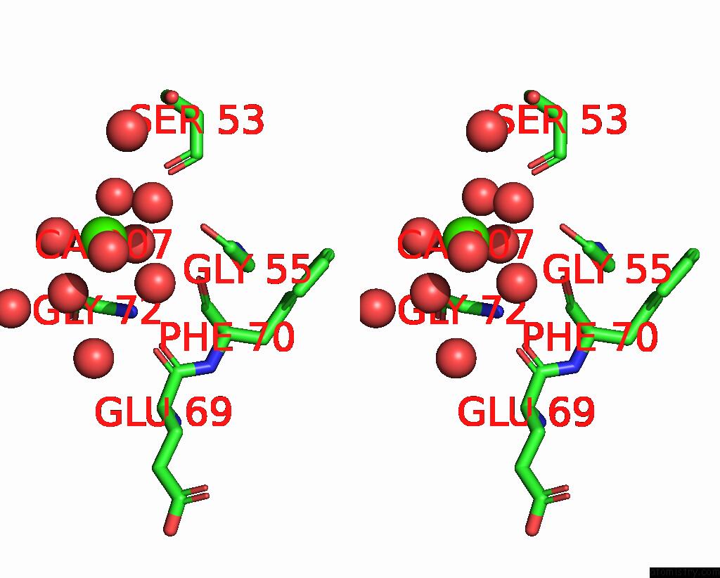
Stereo pair view
|
A full contact list of Calcium with other atoms in the Ca binding
site number 7 of Sucrose-Bound Structure of Marinomonas Primoryensis PA14 Carbohydrate- Binding Domain within 5.0Å range:
probe
|
atom
|
residue
|
distance (Å)
|
B
|
Occ
|
A:Ca207
b:13.2
occ:1.00
|
O
|
A:HOH464
|
2.4
|
11.6
|
1.0
|
O
|
A:HOH350
|
2.4
|
12.8
|
1.0
|
O
|
A:HOH542
|
2.4
|
18.9
|
1.0
|
O
|
A:HOH509
|
2.5
|
18.6
|
1.0
|
O
|
A:HOH436
|
2.5
|
13.1
|
1.0
|
O
|
A:HOH465
|
2.7
|
19.0
|
1.0
|
O
|
A:SER53
|
4.1
|
6.4
|
1.0
|
CA
|
A:GLY72
|
4.3
|
8.2
|
1.0
|
O
|
A:GLY55
|
4.5
|
8.9
|
1.0
|
O
|
A:HOH347
|
4.5
|
20.3
|
1.0
|
O
|
A:HOH444
|
4.6
|
13.3
|
1.0
|
O
|
A:HOH344
|
4.6
|
15.0
|
1.0
|
O
|
A:PHE70
|
4.7
|
5.0
|
1.0
|
O
|
A:GLU69
|
4.8
|
6.6
|
1.0
|
N
|
A:GLY72
|
4.9
|
7.4
|
1.0
|
O
|
A:HOH506
|
4.9
|
35.8
|
1.0
|
|
Reference:
S.Guo,
T.D.R.Vance,
H.Zahiri,
R.Eves,
C.Stevens,
J.H.Hehemann,
S.Vidal-Melgosa,
P.L.Davies.
Structural Basis of Ligand Selectivity By A Bacterial Adhesin Lectin Involved in Multispecies Biofilm Formation. Mbio V. 12 2021.
ISSN: ESSN 2150-7511
PubMed: 33824212
DOI: 10.1128/MBIO.00130-21
Page generated: Tue Jul 16 17:53:38 2024
|