Calcium »
PDB 6x6q-6xqo »
6xqf »
Calcium in PDB 6xqf: Crystal Structure of Sclam E144S Mutant, A Non-Specific Endo-Beta-1, 3(4)-Glucanase From Family GH16, Co-Crystallized with 1,3-Beta-D- Cellotriosyl-Glucose, Presenting A 1,3-Beta-D-Cellobiosyl-Glucose at Active Site
Enzymatic activity of Crystal Structure of Sclam E144S Mutant, A Non-Specific Endo-Beta-1, 3(4)-Glucanase From Family GH16, Co-Crystallized with 1,3-Beta-D- Cellotriosyl-Glucose, Presenting A 1,3-Beta-D-Cellobiosyl-Glucose at Active Site
All present enzymatic activity of Crystal Structure of Sclam E144S Mutant, A Non-Specific Endo-Beta-1, 3(4)-Glucanase From Family GH16, Co-Crystallized with 1,3-Beta-D- Cellotriosyl-Glucose, Presenting A 1,3-Beta-D-Cellobiosyl-Glucose at Active Site:
3.2.1.39;
3.2.1.39;
Protein crystallography data
The structure of Crystal Structure of Sclam E144S Mutant, A Non-Specific Endo-Beta-1, 3(4)-Glucanase From Family GH16, Co-Crystallized with 1,3-Beta-D- Cellotriosyl-Glucose, Presenting A 1,3-Beta-D-Cellobiosyl-Glucose at Active Site, PDB code: 6xqf
was solved by
M.V.Liberato,
F.Squina,
with X-Ray Crystallography technique. A brief refinement statistics is given in the table below:
| Resolution Low / High (Å) | 45.72 / 1.58 |
| Space group | P 21 21 21 |
| Cell size a, b, c (Å), α, β, γ (°) | 38.692, 49.868, 114.474, 90, 90, 90 |
| R / Rfree (%) | 17.1 / 20.9 |
Calcium Binding Sites:
The binding sites of Calcium atom in the Crystal Structure of Sclam E144S Mutant, A Non-Specific Endo-Beta-1, 3(4)-Glucanase From Family GH16, Co-Crystallized with 1,3-Beta-D- Cellotriosyl-Glucose, Presenting A 1,3-Beta-D-Cellobiosyl-Glucose at Active Site
(pdb code 6xqf). This binding sites where shown within
5.0 Angstroms radius around Calcium atom.
In total only one binding site of Calcium was determined in the Crystal Structure of Sclam E144S Mutant, A Non-Specific Endo-Beta-1, 3(4)-Glucanase From Family GH16, Co-Crystallized with 1,3-Beta-D- Cellotriosyl-Glucose, Presenting A 1,3-Beta-D-Cellobiosyl-Glucose at Active Site, PDB code: 6xqf:
In total only one binding site of Calcium was determined in the Crystal Structure of Sclam E144S Mutant, A Non-Specific Endo-Beta-1, 3(4)-Glucanase From Family GH16, Co-Crystallized with 1,3-Beta-D- Cellotriosyl-Glucose, Presenting A 1,3-Beta-D-Cellobiosyl-Glucose at Active Site, PDB code: 6xqf:
Calcium binding site 1 out of 1 in 6xqf
Go back to
Calcium binding site 1 out
of 1 in the Crystal Structure of Sclam E144S Mutant, A Non-Specific Endo-Beta-1, 3(4)-Glucanase From Family GH16, Co-Crystallized with 1,3-Beta-D- Cellotriosyl-Glucose, Presenting A 1,3-Beta-D-Cellobiosyl-Glucose at Active Site
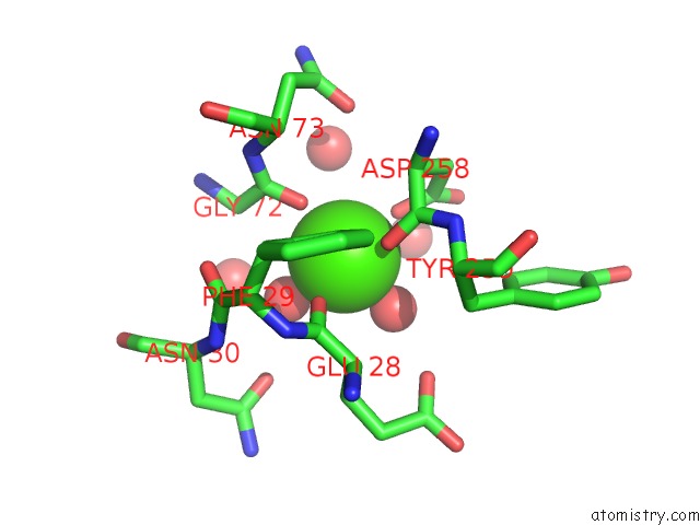
Mono view
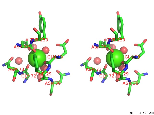
Stereo pair view
Mono view
Stereo pair view
A full contact list of Calcium with other atoms in the Ca binding
site number 1 of Crystal Structure of Sclam E144S Mutant, A Non-Specific Endo-Beta-1, 3(4)-Glucanase From Family GH16, Co-Crystallized with 1,3-Beta-D- Cellotriosyl-Glucose, Presenting A 1,3-Beta-D-Cellobiosyl-Glucose at Active Site within 5.0Å range:
|
Reference:
M.V.Liberato,
E.T.Prates,
A.Bernardes,
J.Fattori,
G.Ematsu,
M.Chinaglia,
E.R.M.Gomes,
A.C.M.Figueira,
I.Polikarpov,
M.Skaf,
F.Squina.
A Structural Basis of the Cleavage Mechanism of Non-Specific Endo-Beta-1,3(4)-Glucanase From Family GH16. To Be Published.
Page generated: Tue Jul 16 18:05:22 2024
Last articles
Zn in 9J0NZn in 9J0O
Zn in 9J0P
Zn in 9FJX
Zn in 9EKB
Zn in 9C0F
Zn in 9CAH
Zn in 9CH0
Zn in 9CH3
Zn in 9CH1