Calcium »
PDB 6xrv-6y41 »
6y3g »
Calcium in PDB 6y3g: Crystal Structure of Phenylalanine Trna From Escherichia Coli
Protein crystallography data
The structure of Crystal Structure of Phenylalanine Trna From Escherichia Coli, PDB code: 6y3g
was solved by
G.Bourgeois,
Y.Mechulam,
E.Schmitt,
with X-Ray Crystallography technique. A brief refinement statistics is given in the table below:
| Resolution Low / High (Å) | 42.98 / 3.10 |
| Space group | P 64 2 2 |
| Cell size a, b, c (Å), α, β, γ (°) | 109.618, 109.618, 138.518, 90, 90, 120 |
| R / Rfree (%) | 21.5 / 23.6 |
Calcium Binding Sites:
The binding sites of Calcium atom in the Crystal Structure of Phenylalanine Trna From Escherichia Coli
(pdb code 6y3g). This binding sites where shown within
5.0 Angstroms radius around Calcium atom.
In total 7 binding sites of Calcium where determined in the Crystal Structure of Phenylalanine Trna From Escherichia Coli, PDB code: 6y3g:
Jump to Calcium binding site number: 1; 2; 3; 4; 5; 6; 7;
In total 7 binding sites of Calcium where determined in the Crystal Structure of Phenylalanine Trna From Escherichia Coli, PDB code: 6y3g:
Jump to Calcium binding site number: 1; 2; 3; 4; 5; 6; 7;
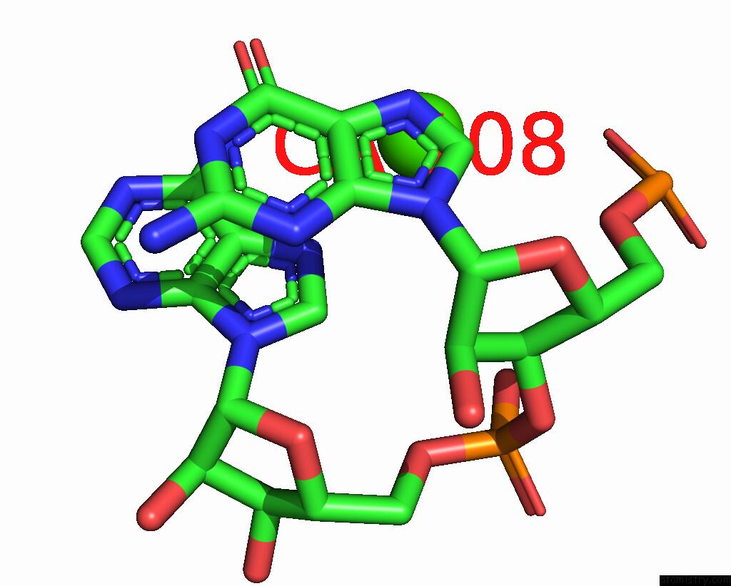
Calcium binding site 1 out of 7 in 6y3g
Go back to
Calcium binding site 1 out
of 7 in the Crystal Structure of Phenylalanine Trna From Escherichia Coli

Mono view

Stereo pair view
Mono view
Stereo pair view
A full contact list of Calcium with other atoms in the Ca binding
site number 1 of Crystal Structure of Phenylalanine Trna From Escherichia Coli within 5.0Å range:
|
Calcium binding site 2 out of 7 in 6y3g
Go back to
Calcium binding site 2 out
of 7 in the Crystal Structure of Phenylalanine Trna From Escherichia Coli

Mono view

Stereo pair view
Mono view
Stereo pair view
A full contact list of Calcium with other atoms in the Ca binding
site number 2 of Crystal Structure of Phenylalanine Trna From Escherichia Coli within 5.0Å range:
|
Calcium binding site 3 out of 7 in 6y3g
Go back to
Calcium binding site 3 out
of 7 in the Crystal Structure of Phenylalanine Trna From Escherichia Coli

Mono view

Stereo pair view
Mono view
Stereo pair view
A full contact list of Calcium with other atoms in the Ca binding
site number 3 of Crystal Structure of Phenylalanine Trna From Escherichia Coli within 5.0Å range:
|
Calcium binding site 4 out of 7 in 6y3g
Go back to
Calcium binding site 4 out
of 7 in the Crystal Structure of Phenylalanine Trna From Escherichia Coli
Mono view

Stereo pair view
Mono view
Stereo pair view
A full contact list of Calcium with other atoms in the Ca binding
site number 4 of Crystal Structure of Phenylalanine Trna From Escherichia Coli within 5.0Å range:
|
Calcium binding site 5 out of 7 in 6y3g
Go back to
Calcium binding site 5 out
of 7 in the Crystal Structure of Phenylalanine Trna From Escherichia Coli

Mono view

Stereo pair view
Mono view
Stereo pair view
A full contact list of Calcium with other atoms in the Ca binding
site number 5 of Crystal Structure of Phenylalanine Trna From Escherichia Coli within 5.0Å range:
|
Calcium binding site 6 out of 7 in 6y3g
Go back to
Calcium binding site 6 out
of 7 in the Crystal Structure of Phenylalanine Trna From Escherichia Coli

Mono view

Stereo pair view
Mono view
Stereo pair view
A full contact list of Calcium with other atoms in the Ca binding
site number 6 of Crystal Structure of Phenylalanine Trna From Escherichia Coli within 5.0Å range:
|
Calcium binding site 7 out of 7 in 6y3g
Go back to
Calcium binding site 7 out
of 7 in the Crystal Structure of Phenylalanine Trna From Escherichia Coli

Mono view

Stereo pair view
Mono view
Stereo pair view
A full contact list of Calcium with other atoms in the Ca binding
site number 7 of Crystal Structure of Phenylalanine Trna From Escherichia Coli within 5.0Å range:
|
Reference:
G.Bourgeois,
J.Seguin,
M.Babin,
M.Gondry,
Y.Mechulam,
E.Schmitt.
Structural Basis of the Interaction Between Cyclodipeptide Synthases and Aminoacylated Trna Substrates. Rna V. 26 1589 2020.
ISSN: ESSN 1469-9001
PubMed: 32680846
DOI: 10.1261/RNA.075184.120
Page generated: Tue Jul 16 18:23:54 2024
ISSN: ESSN 1469-9001
PubMed: 32680846
DOI: 10.1261/RNA.075184.120
Last articles
Zn in 9J0NZn in 9J0O
Zn in 9J0P
Zn in 9FJX
Zn in 9EKB
Zn in 9C0F
Zn in 9CAH
Zn in 9CH0
Zn in 9CH3
Zn in 9CH1