Calcium »
PDB 6y43-6ycw »
6y8z »
Calcium in PDB 6y8z: Structure of Baltic Herring (Clupea Harengus) Phosphoglucomutase 5 (PGM5)
Protein crystallography data
The structure of Structure of Baltic Herring (Clupea Harengus) Phosphoglucomutase 5 (PGM5), PDB code: 6y8z
was solved by
R.Gustafsson,
U.Eckhard,
M.Selmer,
with X-Ray Crystallography technique. A brief refinement statistics is given in the table below:
| Resolution Low / High (Å) | 44.93 / 2.05 |
| Space group | P 2 21 21 |
| Cell size a, b, c (Å), α, β, γ (°) | 47.200, 94.210, 146.690, 90.00, 90.00, 90.00 |
| R / Rfree (%) | 18.7 / 23.6 |
Other elements in 6y8z:
The structure of Structure of Baltic Herring (Clupea Harengus) Phosphoglucomutase 5 (PGM5) also contains other interesting chemical elements:
| Nickel | (Ni) | 1 atom |
| Sodium | (Na) | 4 atoms |
Calcium Binding Sites:
The binding sites of Calcium atom in the Structure of Baltic Herring (Clupea Harengus) Phosphoglucomutase 5 (PGM5)
(pdb code 6y8z). This binding sites where shown within
5.0 Angstroms radius around Calcium atom.
In total only one binding site of Calcium was determined in the Structure of Baltic Herring (Clupea Harengus) Phosphoglucomutase 5 (PGM5), PDB code: 6y8z:
In total only one binding site of Calcium was determined in the Structure of Baltic Herring (Clupea Harengus) Phosphoglucomutase 5 (PGM5), PDB code: 6y8z:
Calcium binding site 1 out of 1 in 6y8z
Go back to
Calcium binding site 1 out
of 1 in the Structure of Baltic Herring (Clupea Harengus) Phosphoglucomutase 5 (PGM5)
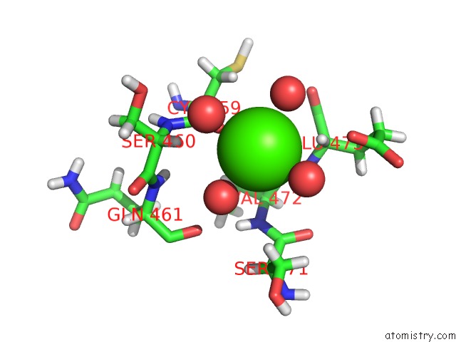
Mono view
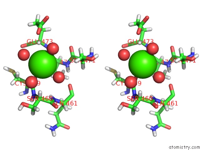
Stereo pair view
Mono view
Stereo pair view
A full contact list of Calcium with other atoms in the Ca binding
site number 1 of Structure of Baltic Herring (Clupea Harengus) Phosphoglucomutase 5 (PGM5) within 5.0Å range:
|
Reference:
R.Gustafsson,
U.Eckhard,
W.Ye,
E.D.Enbody,
M.Pettersson,
P.Jemth,
L.Andersson,
M.Selmer.
Structure and Characterization of Phosphoglucomutase 5 From Atlantic and Baltic Herring-An Inactive Enzyme with Intact Substrate Binding Biomolecules 2020.
ISSN: ESSN 2218-273X
DOI: 10.3390/BIOM10121631
Page generated: Thu Jul 18 21:48:14 2024
ISSN: ESSN 2218-273X
DOI: 10.3390/BIOM10121631
Last articles
Zn in 9MJ5Zn in 9HNW
Zn in 9G0L
Zn in 9FNE
Zn in 9DZN
Zn in 9E0I
Zn in 9D32
Zn in 9DAK
Zn in 8ZXC
Zn in 8ZUF