Calcium »
PDB 6yd4-6ywn »
6yii »
Calcium in PDB 6yii: Crystal Structure of A Class III Adenylyl Cyclase-Like Atp-Binding Protein From Pseudomonas Aeruginosa
Protein crystallography data
The structure of Crystal Structure of A Class III Adenylyl Cyclase-Like Atp-Binding Protein From Pseudomonas Aeruginosa, PDB code: 6yii
was solved by
S.Moniot,
C.Steegborn,
with X-Ray Crystallography technique. A brief refinement statistics is given in the table below:
| Resolution Low / High (Å) | 30.54 / 1.44 |
| Space group | P 21 21 21 |
| Cell size a, b, c (Å), α, β, γ (°) | 62.400, 74.680, 149.680, 90.00, 90.00, 90.00 |
| R / Rfree (%) | 13.1 / 14.7 |
Other elements in 6yii:
The structure of Crystal Structure of A Class III Adenylyl Cyclase-Like Atp-Binding Protein From Pseudomonas Aeruginosa also contains other interesting chemical elements:
| Magnesium | (Mg) | 1 atom |
| Manganese | (Mn) | 1 atom |
| Sodium | (Na) | 3 atoms |
Calcium Binding Sites:
The binding sites of Calcium atom in the Crystal Structure of A Class III Adenylyl Cyclase-Like Atp-Binding Protein From Pseudomonas Aeruginosa
(pdb code 6yii). This binding sites where shown within
5.0 Angstroms radius around Calcium atom.
In total only one binding site of Calcium was determined in the Crystal Structure of A Class III Adenylyl Cyclase-Like Atp-Binding Protein From Pseudomonas Aeruginosa, PDB code: 6yii:
In total only one binding site of Calcium was determined in the Crystal Structure of A Class III Adenylyl Cyclase-Like Atp-Binding Protein From Pseudomonas Aeruginosa, PDB code: 6yii:
Calcium binding site 1 out of 1 in 6yii
Go back to
Calcium binding site 1 out
of 1 in the Crystal Structure of A Class III Adenylyl Cyclase-Like Atp-Binding Protein From Pseudomonas Aeruginosa
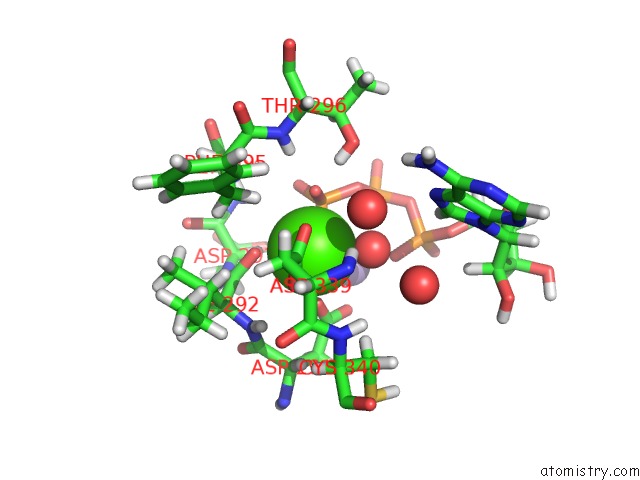
Mono view
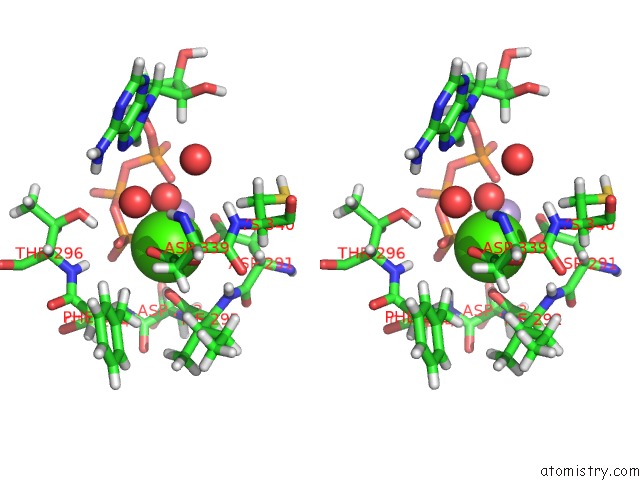
Stereo pair view
Mono view
Stereo pair view
A full contact list of Calcium with other atoms in the Ca binding
site number 1 of Crystal Structure of A Class III Adenylyl Cyclase-Like Atp-Binding Protein From Pseudomonas Aeruginosa within 5.0Å range:
|
Reference:
J.Linder,
E.Hupfeld,
M.Weyand,
C.Steegborn,
S.Moniot.
Crystal Structure of A Class III Adenylyl Cyclase-Like Atp-Binding Protein From Pseudomonas Aeruginosa. J.Struct.Biol. 07534 2020.
ISSN: ESSN 1095-8657
PubMed: 32454240
DOI: 10.1016/J.JSB.2020.107534
Page generated: Thu Jul 18 22:23:31 2024
ISSN: ESSN 1095-8657
PubMed: 32454240
DOI: 10.1016/J.JSB.2020.107534
Last articles
Zn in 9J0NZn in 9J0O
Zn in 9J0P
Zn in 9FJX
Zn in 9EKB
Zn in 9C0F
Zn in 9CAH
Zn in 9CH0
Zn in 9CH3
Zn in 9CH1