Calcium in PDB 6ype: Crystal Structure of the Human Neuronal Pentraxin 1 (NP1) Pentraxin (Ptx) Domain.
Protein crystallography data
The structure of Crystal Structure of the Human Neuronal Pentraxin 1 (NP1) Pentraxin (Ptx) Domain., PDB code: 6ype
was solved by
J.Elegheert,
A.J.Clayton,
A.R.Aricescu,
with X-Ray Crystallography technique. A brief refinement statistics is given in the table below:
Resolution Low / High (Å)
|
45.69 /
1.45
|
Space group
|
C 1 2 1
|
Cell size a, b, c (Å), α, β, γ (°)
|
121.220,
36.060,
92.170,
90.00,
97.47,
90.00
|
R / Rfree (%)
|
15.3 /
17.8
|
Other elements in 6ype:
The structure of Crystal Structure of the Human Neuronal Pentraxin 1 (NP1) Pentraxin (Ptx) Domain. also contains other interesting chemical elements:
Calcium Binding Sites:
The binding sites of Calcium atom in the Crystal Structure of the Human Neuronal Pentraxin 1 (NP1) Pentraxin (Ptx) Domain.
(pdb code 6ype). This binding sites where shown within
5.0 Angstroms radius around Calcium atom.
In total 4 binding sites of Calcium where determined in the
Crystal Structure of the Human Neuronal Pentraxin 1 (NP1) Pentraxin (Ptx) Domain., PDB code: 6ype:
Jump to Calcium binding site number:
1;
2;
3;
4;
Calcium binding site 1 out
of 4 in 6ype
Go back to
Calcium Binding Sites List in 6ype
Calcium binding site 1 out
of 4 in the Crystal Structure of the Human Neuronal Pentraxin 1 (NP1) Pentraxin (Ptx) Domain.
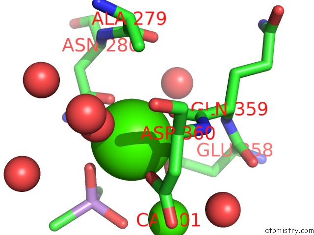
Mono view
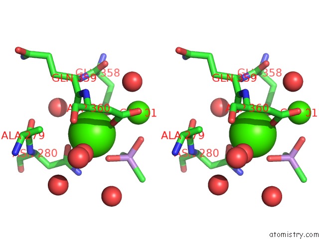
Stereo pair view
|
A full contact list of Calcium with other atoms in the Ca binding
site number 1 of Crystal Structure of the Human Neuronal Pentraxin 1 (NP1) Pentraxin (Ptx) Domain. within 5.0Å range:
probe
|
atom
|
residue
|
distance (Å)
|
B
|
Occ
|
A:Ca500
b:9.0
occ:1.00
|
O
|
A:GLN359
|
2.3
|
8.8
|
1.0
|
O2
|
A:CAC502
|
2.3
|
21.0
|
1.0
|
OD1
|
A:ASN280
|
2.3
|
10.0
|
1.0
|
OE1
|
A:GLU358
|
2.4
|
9.8
|
1.0
|
OD1
|
A:ASP360
|
2.4
|
9.6
|
1.0
|
O
|
A:HOH746
|
2.4
|
13.1
|
1.0
|
OE2
|
A:GLU358
|
2.6
|
9.6
|
1.0
|
CD
|
A:GLU358
|
2.8
|
12.7
|
1.0
|
CG
|
A:ASN280
|
3.3
|
7.7
|
1.0
|
C
|
A:GLN359
|
3.3
|
9.6
|
1.0
|
ND2
|
A:ASN280
|
3.6
|
10.2
|
1.0
|
CG
|
A:ASP360
|
3.6
|
9.5
|
1.0
|
AS
|
A:CAC502
|
3.6
|
20.5
|
1.0
|
O1
|
A:CAC502
|
3.8
|
21.0
|
1.0
|
CA
|
A:ASP360
|
3.8
|
7.7
|
1.0
|
CA
|
A:CA501
|
3.8
|
9.0
|
1.0
|
N
|
A:ASP360
|
3.9
|
8.8
|
1.0
|
O
|
A:HOH675
|
4.0
|
9.1
|
1.0
|
CB
|
A:ASP360
|
4.3
|
10.9
|
1.0
|
CG
|
A:GLU358
|
4.3
|
8.6
|
1.0
|
C1
|
A:CAC502
|
4.4
|
20.7
|
1.0
|
N
|
A:GLN359
|
4.4
|
8.7
|
1.0
|
CA
|
A:GLN359
|
4.5
|
8.9
|
1.0
|
CB
|
A:ALA279
|
4.5
|
8.8
|
1.0
|
OD2
|
A:ASP360
|
4.5
|
11.5
|
1.0
|
CB
|
A:ASN280
|
4.7
|
7.8
|
1.0
|
O
|
A:ALA279
|
4.7
|
9.3
|
1.0
|
O
|
A:HOH669
|
4.7
|
19.8
|
1.0
|
C
|
A:ALA279
|
4.8
|
8.8
|
1.0
|
O
|
A:HOH654
|
4.8
|
29.1
|
1.0
|
N
|
A:ASN280
|
4.9
|
9.6
|
1.0
|
O
|
A:HOH763
|
4.9
|
35.5
|
1.0
|
O
|
A:HOH624
|
5.0
|
10.4
|
1.0
|
|
Calcium binding site 2 out
of 4 in 6ype
Go back to
Calcium Binding Sites List in 6ype
Calcium binding site 2 out
of 4 in the Crystal Structure of the Human Neuronal Pentraxin 1 (NP1) Pentraxin (Ptx) Domain.
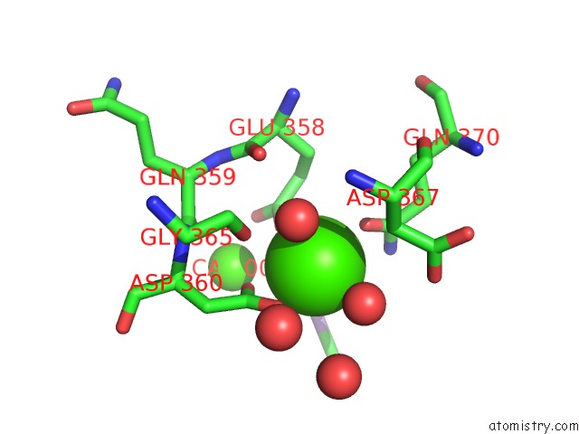
Mono view
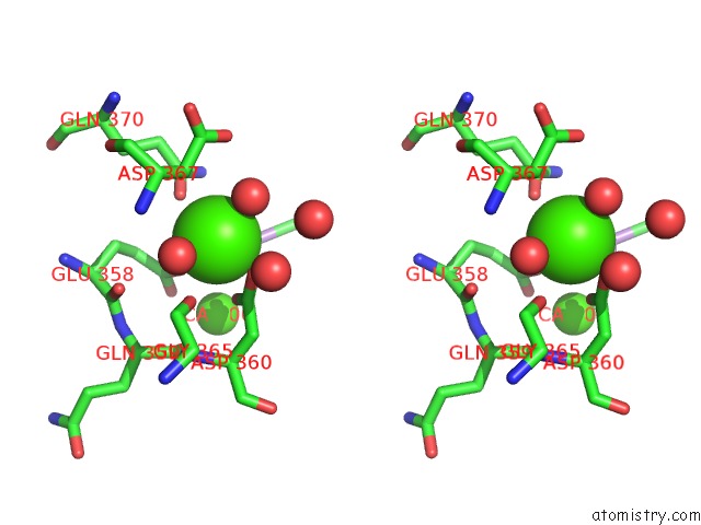
Stereo pair view
|
A full contact list of Calcium with other atoms in the Ca binding
site number 2 of Crystal Structure of the Human Neuronal Pentraxin 1 (NP1) Pentraxin (Ptx) Domain. within 5.0Å range:
probe
|
atom
|
residue
|
distance (Å)
|
B
|
Occ
|
A:Ca501
b:9.0
occ:1.00
|
O1
|
A:CAC502
|
2.2
|
21.0
|
1.0
|
OE2
|
A:GLU358
|
2.3
|
9.6
|
1.0
|
OE1
|
A:GLN370
|
2.4
|
9.4
|
1.0
|
O
|
A:HOH635
|
2.4
|
14.5
|
1.0
|
O
|
A:HOH624
|
2.4
|
10.4
|
1.0
|
OD2
|
A:ASP360
|
2.5
|
11.5
|
1.0
|
OD1
|
A:ASP360
|
2.6
|
9.6
|
1.0
|
CG
|
A:ASP360
|
2.9
|
9.5
|
1.0
|
CD
|
A:GLN370
|
3.3
|
12.7
|
1.0
|
CD
|
A:GLU358
|
3.5
|
12.7
|
1.0
|
NE2
|
A:GLN370
|
3.6
|
13.0
|
1.0
|
AS
|
A:CAC502
|
3.7
|
20.5
|
1.0
|
CA
|
A:CA500
|
3.8
|
9.0
|
1.0
|
CB
|
A:ASP367
|
4.1
|
9.3
|
1.0
|
O2
|
A:CAC502
|
4.2
|
21.0
|
1.0
|
CG
|
A:GLU358
|
4.3
|
8.6
|
1.0
|
CB
|
A:ASP360
|
4.3
|
10.9
|
1.0
|
CB
|
A:GLU358
|
4.3
|
8.2
|
1.0
|
OE1
|
A:GLU358
|
4.4
|
9.8
|
1.0
|
O
|
A:GLY365
|
4.5
|
13.5
|
1.0
|
OD2
|
A:ASP367
|
4.5
|
15.5
|
1.0
|
O
|
A:GLU358
|
4.5
|
10.3
|
1.0
|
O
|
A:HOH767
|
4.6
|
44.8
|
1.0
|
CG
|
A:GLN370
|
4.7
|
8.8
|
1.0
|
O
|
A:GLN359
|
4.7
|
8.8
|
1.0
|
C1
|
A:CAC502
|
4.7
|
20.7
|
1.0
|
C
|
A:GLU358
|
4.8
|
8.9
|
1.0
|
C
|
A:GLN359
|
4.8
|
9.6
|
1.0
|
N
|
A:ASP360
|
4.8
|
8.8
|
1.0
|
O
|
A:HOH642
|
4.9
|
19.5
|
1.0
|
CG
|
A:ASP367
|
4.9
|
16.7
|
1.0
|
CA
|
A:ASP360
|
5.0
|
7.7
|
1.0
|
|
Calcium binding site 3 out
of 4 in 6ype
Go back to
Calcium Binding Sites List in 6ype
Calcium binding site 3 out
of 4 in the Crystal Structure of the Human Neuronal Pentraxin 1 (NP1) Pentraxin (Ptx) Domain.
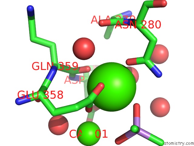
Mono view
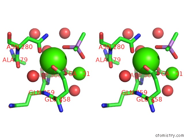
Stereo pair view
|
A full contact list of Calcium with other atoms in the Ca binding
site number 3 of Crystal Structure of the Human Neuronal Pentraxin 1 (NP1) Pentraxin (Ptx) Domain. within 5.0Å range:
probe
|
atom
|
residue
|
distance (Å)
|
B
|
Occ
|
B:Ca500
b:9.6
occ:1.00
|
O
|
B:GLN359
|
2.3
|
10.7
|
1.0
|
O1
|
B:CAC502
|
2.3
|
16.0
|
1.0
|
OD1
|
B:ASP360
|
2.3
|
11.0
|
1.0
|
OD1
|
B:ASN280
|
2.4
|
8.7
|
1.0
|
O
|
B:HOH748
|
2.4
|
11.5
|
1.0
|
OE2
|
B:GLU358
|
2.4
|
9.7
|
1.0
|
OE1
|
B:GLU358
|
2.5
|
9.9
|
1.0
|
CD
|
B:GLU358
|
2.8
|
8.6
|
1.0
|
C
|
B:GLN359
|
3.3
|
9.8
|
1.0
|
CG
|
B:ASN280
|
3.4
|
9.3
|
1.0
|
CG
|
B:ASP360
|
3.6
|
8.8
|
1.0
|
AS
|
B:CAC502
|
3.6
|
15.8
|
1.0
|
ND2
|
B:ASN280
|
3.7
|
10.8
|
1.0
|
CA
|
B:ASP360
|
3.8
|
9.5
|
1.0
|
CA
|
B:CA501
|
3.8
|
11.1
|
1.0
|
N
|
B:ASP360
|
3.9
|
9.3
|
1.0
|
O2
|
B:CAC502
|
3.9
|
15.9
|
1.0
|
O
|
B:HOH677
|
4.0
|
8.5
|
1.0
|
CB
|
B:ASP360
|
4.3
|
13.2
|
1.0
|
CG
|
B:GLU358
|
4.3
|
10.4
|
1.0
|
C2
|
B:CAC502
|
4.3
|
15.8
|
1.0
|
O
|
B:HOH700
|
4.4
|
24.7
|
1.0
|
N
|
B:GLN359
|
4.4
|
7.0
|
1.0
|
CA
|
B:GLN359
|
4.5
|
6.5
|
1.0
|
OD2
|
B:ASP360
|
4.5
|
10.8
|
1.0
|
CB
|
B:ALA279
|
4.6
|
10.2
|
1.0
|
O
|
B:HOH647
|
4.6
|
35.6
|
1.0
|
O
|
B:ALA279
|
4.7
|
8.6
|
1.0
|
O
|
B:HOH683
|
4.7
|
15.2
|
1.0
|
CB
|
B:ASN280
|
4.7
|
7.0
|
1.0
|
C
|
B:ALA279
|
4.8
|
8.5
|
1.0
|
N
|
B:ASN280
|
4.9
|
7.3
|
1.0
|
O
|
B:HOH646
|
4.9
|
9.2
|
1.0
|
C
|
B:GLU358
|
5.0
|
7.1
|
1.0
|
|
Calcium binding site 4 out
of 4 in 6ype
Go back to
Calcium Binding Sites List in 6ype
Calcium binding site 4 out
of 4 in the Crystal Structure of the Human Neuronal Pentraxin 1 (NP1) Pentraxin (Ptx) Domain.
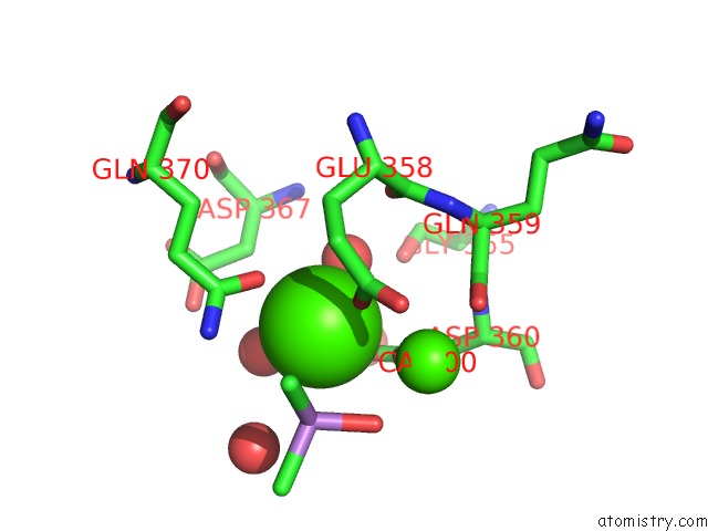
Mono view
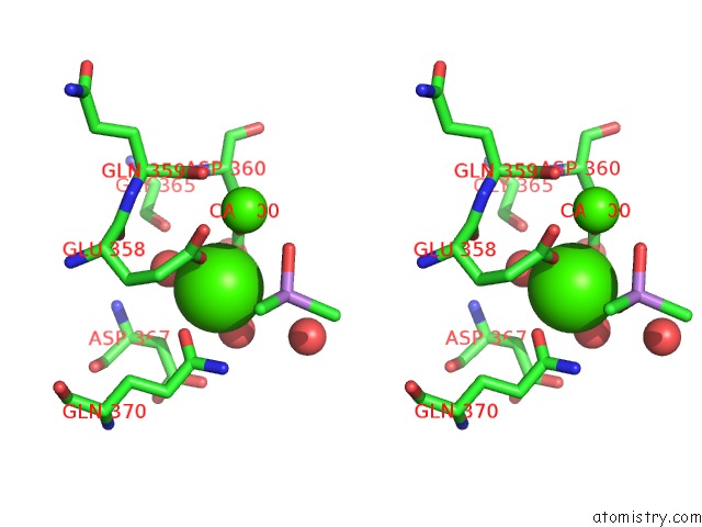
Stereo pair view
|
A full contact list of Calcium with other atoms in the Ca binding
site number 4 of Crystal Structure of the Human Neuronal Pentraxin 1 (NP1) Pentraxin (Ptx) Domain. within 5.0Å range:
probe
|
atom
|
residue
|
distance (Å)
|
B
|
Occ
|
B:Ca501
b:11.1
occ:1.00
|
O2
|
B:CAC502
|
2.3
|
15.9
|
1.0
|
OE1
|
B:GLU358
|
2.3
|
9.9
|
1.0
|
O
|
B:HOH640
|
2.4
|
12.9
|
1.0
|
O
|
B:HOH646
|
2.4
|
9.2
|
1.0
|
OE1
|
B:GLN370
|
2.4
|
10.8
|
1.0
|
OD1
|
B:ASP360
|
2.5
|
11.0
|
1.0
|
OD2
|
B:ASP360
|
2.6
|
10.8
|
1.0
|
CG
|
B:ASP360
|
2.9
|
8.8
|
1.0
|
CD
|
B:GLN370
|
3.3
|
11.9
|
1.0
|
CD
|
B:GLU358
|
3.5
|
8.6
|
1.0
|
NE2
|
B:GLN370
|
3.6
|
14.2
|
1.0
|
AS
|
B:CAC502
|
3.7
|
15.8
|
1.0
|
CA
|
B:CA500
|
3.8
|
9.6
|
1.0
|
O
|
B:HOH638
|
4.1
|
21.9
|
1.0
|
O1
|
B:CAC502
|
4.1
|
16.0
|
1.0
|
CB
|
B:ASP367
|
4.1
|
11.3
|
1.0
|
CG
|
B:GLU358
|
4.3
|
10.4
|
1.0
|
CB
|
B:ASP360
|
4.3
|
13.2
|
1.0
|
CB
|
B:GLU358
|
4.3
|
9.6
|
1.0
|
OE2
|
B:GLU358
|
4.4
|
9.7
|
1.0
|
OD2
|
B:ASP367
|
4.5
|
16.0
|
1.0
|
O
|
B:GLU358
|
4.5
|
7.9
|
1.0
|
O
|
B:GLY365
|
4.6
|
11.7
|
1.0
|
C2
|
B:CAC502
|
4.6
|
15.8
|
1.0
|
CG
|
B:GLN370
|
4.7
|
10.4
|
1.0
|
O
|
B:GLN359
|
4.8
|
10.7
|
1.0
|
C
|
B:GLU358
|
4.8
|
7.1
|
1.0
|
C
|
B:GLN359
|
4.8
|
9.8
|
1.0
|
N
|
B:ASP360
|
4.8
|
9.3
|
1.0
|
CG
|
B:ASP367
|
4.9
|
17.9
|
1.0
|
O
|
B:HOH635
|
4.9
|
20.9
|
1.0
|
CA
|
B:ASP360
|
5.0
|
9.5
|
1.0
|
|
Reference:
K.Suzuki,
J.Elegheert,
I.Song,
H.Sasakura,
O.Senkov,
W.Kakegawa,
A.J.Clayton,
V.T.Chang,
M.Ferrer-Ferrer,
E.Miura,
R.Kaushik,
M.Ikeno,
Y.Morioka,
Y.Takeuchi,
T.Shimada,
S.Otsuka,
S.Stoyanov,
M.Watanabe,
K.Takeuchi,
A.Dityatev,
A.R.Aricescu,
M.Yuzaki.
A Synthetic Synaptic Organizer Protein Restores Glutamatergic Neuronal Circuits. To Be Published.
Page generated: Thu Jul 18 22:28:07 2024
|