Calcium »
PDB 6yxh-6zqx »
6zdy »
Calcium in PDB 6zdy: Crystal Structure of Wt Murine S100A9 Bound to Calcium and Zinc
Protein crystallography data
The structure of Crystal Structure of Wt Murine S100A9 Bound to Calcium and Zinc, PDB code: 6zdy
was solved by
L.Yatime,
with X-Ray Crystallography technique. A brief refinement statistics is given in the table below:
| Resolution Low / High (Å) | 43.49 / 1.45 |
| Space group | C 1 2 1 |
| Cell size a, b, c (Å), α, β, γ (°) | 72.84, 40.62, 53.09, 90, 125, 90 |
| R / Rfree (%) | 17.6 / 20 |
Other elements in 6zdy:
The structure of Crystal Structure of Wt Murine S100A9 Bound to Calcium and Zinc also contains other interesting chemical elements:
| Zinc | (Zn) | 2 atoms |
Calcium Binding Sites:
The binding sites of Calcium atom in the Crystal Structure of Wt Murine S100A9 Bound to Calcium and Zinc
(pdb code 6zdy). This binding sites where shown within
5.0 Angstroms radius around Calcium atom.
In total 2 binding sites of Calcium where determined in the Crystal Structure of Wt Murine S100A9 Bound to Calcium and Zinc, PDB code: 6zdy:
Jump to Calcium binding site number: 1; 2;
In total 2 binding sites of Calcium where determined in the Crystal Structure of Wt Murine S100A9 Bound to Calcium and Zinc, PDB code: 6zdy:
Jump to Calcium binding site number: 1; 2;
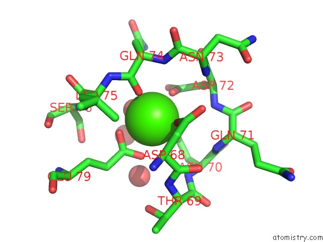
Calcium binding site 1 out of 2 in 6zdy
Go back to
Calcium binding site 1 out
of 2 in the Crystal Structure of Wt Murine S100A9 Bound to Calcium and Zinc

Mono view

Stereo pair view
Mono view
Stereo pair view
A full contact list of Calcium with other atoms in the Ca binding
site number 1 of Crystal Structure of Wt Murine S100A9 Bound to Calcium and Zinc within 5.0Å range:
|
Calcium binding site 2 out of 2 in 6zdy
Go back to
Calcium binding site 2 out
of 2 in the Crystal Structure of Wt Murine S100A9 Bound to Calcium and Zinc
Mono view
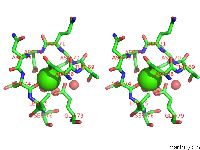
Stereo pair view
Mono view
Stereo pair view
A full contact list of Calcium with other atoms in the Ca binding
site number 2 of Crystal Structure of Wt Murine S100A9 Bound to Calcium and Zinc within 5.0Å range:
|
Reference:
L.Signor,
T.Paris,
C.Mas,
A.Picard,
G.Lutfalla,
E.Boeri Erba,
L.Yatime.
Divalent Cations Influence the Dimerization Mode of Murine S100A9 Protein By Modulating Its Disulfide Bond Pattern. J.Struct.Biol. V. 213 07689 2020.
ISSN: ESSN 1095-8657
PubMed: 33359632
DOI: 10.1016/J.JSB.2020.107689
Page generated: Thu Jul 18 22:39:31 2024
ISSN: ESSN 1095-8657
PubMed: 33359632
DOI: 10.1016/J.JSB.2020.107689
Last articles
Zn in 9J0NZn in 9J0O
Zn in 9J0P
Zn in 9FJX
Zn in 9EKB
Zn in 9C0F
Zn in 9CAH
Zn in 9CH0
Zn in 9CH3
Zn in 9CH1