Calcium »
PDB 6zr0-7aq1 »
7a1r »
Calcium in PDB 7a1r: Crystal Structure of the C2B Domain of Trypanosoma Brucei Extended Synaptotagmin (E-Syt)
Protein crystallography data
The structure of Crystal Structure of the C2B Domain of Trypanosoma Brucei Extended Synaptotagmin (E-Syt), PDB code: 7a1r
was solved by
G.Dong,
with X-Ray Crystallography technique. A brief refinement statistics is given in the table below:
| Resolution Low / High (Å) | 19.86 / 1.50 |
| Space group | P 21 21 21 |
| Cell size a, b, c (Å), α, β, γ (°) | 54.908, 57.532, 84.57, 90, 90, 90 |
| R / Rfree (%) | 17 / 19.1 |
Other elements in 7a1r:
The structure of Crystal Structure of the C2B Domain of Trypanosoma Brucei Extended Synaptotagmin (E-Syt) also contains other interesting chemical elements:
| Chlorine | (Cl) | 7 atoms |
| Sodium | (Na) | 1 atom |
Calcium Binding Sites:
The binding sites of Calcium atom in the Crystal Structure of the C2B Domain of Trypanosoma Brucei Extended Synaptotagmin (E-Syt)
(pdb code 7a1r). This binding sites where shown within
5.0 Angstroms radius around Calcium atom.
In total 4 binding sites of Calcium where determined in the Crystal Structure of the C2B Domain of Trypanosoma Brucei Extended Synaptotagmin (E-Syt), PDB code: 7a1r:
Jump to Calcium binding site number: 1; 2; 3; 4;
In total 4 binding sites of Calcium where determined in the Crystal Structure of the C2B Domain of Trypanosoma Brucei Extended Synaptotagmin (E-Syt), PDB code: 7a1r:
Jump to Calcium binding site number: 1; 2; 3; 4;
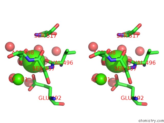
Calcium binding site 1 out of 4 in 7a1r
Go back to
Calcium binding site 1 out
of 4 in the Crystal Structure of the C2B Domain of Trypanosoma Brucei Extended Synaptotagmin (E-Syt)

Mono view

Stereo pair view
Mono view
Stereo pair view
A full contact list of Calcium with other atoms in the Ca binding
site number 1 of Crystal Structure of the C2B Domain of Trypanosoma Brucei Extended Synaptotagmin (E-Syt) within 5.0Å range:
|
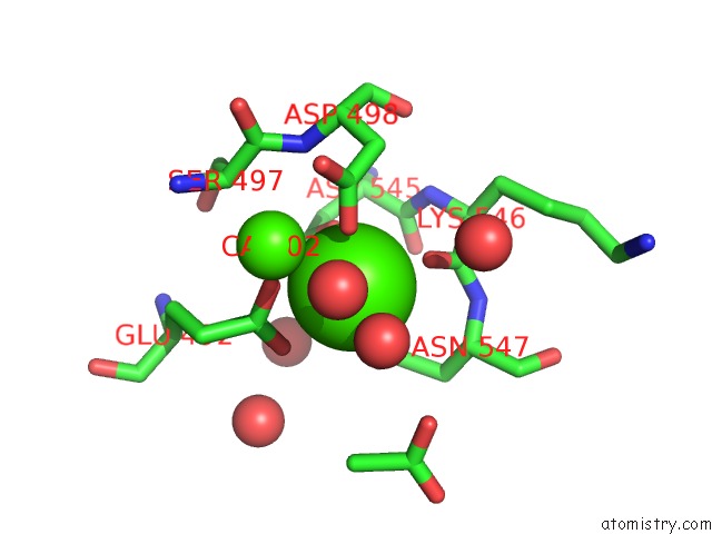
Calcium binding site 2 out of 4 in 7a1r
Go back to
Calcium binding site 2 out
of 4 in the Crystal Structure of the C2B Domain of Trypanosoma Brucei Extended Synaptotagmin (E-Syt)

Mono view
Stereo pair view
Mono view
Stereo pair view
A full contact list of Calcium with other atoms in the Ca binding
site number 2 of Crystal Structure of the C2B Domain of Trypanosoma Brucei Extended Synaptotagmin (E-Syt) within 5.0Å range:
|
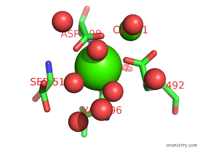
Calcium binding site 3 out of 4 in 7a1r
Go back to
Calcium binding site 3 out
of 4 in the Crystal Structure of the C2B Domain of Trypanosoma Brucei Extended Synaptotagmin (E-Syt)
Mono view

Stereo pair view
Mono view
Stereo pair view
A full contact list of Calcium with other atoms in the Ca binding
site number 3 of Crystal Structure of the C2B Domain of Trypanosoma Brucei Extended Synaptotagmin (E-Syt) within 5.0Å range:
|
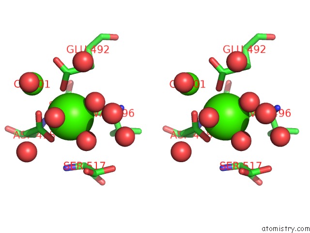
Calcium binding site 4 out of 4 in 7a1r
Go back to
Calcium binding site 4 out
of 4 in the Crystal Structure of the C2B Domain of Trypanosoma Brucei Extended Synaptotagmin (E-Syt)
Mono view
Stereo pair view
Mono view
Stereo pair view
A full contact list of Calcium with other atoms in the Ca binding
site number 4 of Crystal Structure of the C2B Domain of Trypanosoma Brucei Extended Synaptotagmin (E-Syt) within 5.0Å range:
|
Reference:
E.Stepinac,
N.Landrein,
D.Skwarzynska,
P.Wojcik,
J.Lesigang,
I.Lucic,
C.Y.He,
M.Bonhivers,
D.R.Robinson,
G.Dong.
Structural Studies of the Shortest Extended Synaptotagmin with Only Two C2 Domains From Trypanosoma Brucei . Iscience V. 24 02422 2021.
ISSN: ESSN 2589-0042
PubMed: 33997700
DOI: 10.1016/J.ISCI.2021.102422
Page generated: Wed Jul 9 20:39:53 2025
ISSN: ESSN 2589-0042
PubMed: 33997700
DOI: 10.1016/J.ISCI.2021.102422
Last articles
F in 7M2NF in 7M4V
F in 7M4U
F in 7M3F
F in 7M3S
F in 7M4T
F in 7M4P
F in 7M04
F in 7M2L
F in 7M2O