Calcium »
PDB 6zqy-7aq0 »
7ag5 »
Calcium in PDB 7ag5: Structure of the Laspartomycin C Double Mutant G4D D-Allo-THR9D-Dap in Complex with Geranyl Phosphate
Protein crystallography data
The structure of Structure of the Laspartomycin C Double Mutant G4D D-Allo-THR9D-Dap in Complex with Geranyl Phosphate, PDB code: 7ag5
was solved by
M.R.Zeronian,
N.M.Pearce,
T.M.Wood,
N.I.Martin,
B.J.C.Janssen,
with X-Ray Crystallography technique. A brief refinement statistics is given in the table below:
| Resolution Low / High (Å) | 35.01 / 1.04 |
| Space group | P 6 2 2 |
| Cell size a, b, c (Å), α, β, γ (°) | 40.428, 40.428, 31.033, 90, 90, 120 |
| R / Rfree (%) | 12 / 14.3 |
Calcium Binding Sites:
The binding sites of Calcium atom in the Structure of the Laspartomycin C Double Mutant G4D D-Allo-THR9D-Dap in Complex with Geranyl Phosphate
(pdb code 7ag5). This binding sites where shown within
5.0 Angstroms radius around Calcium atom.
In total 2 binding sites of Calcium where determined in the Structure of the Laspartomycin C Double Mutant G4D D-Allo-THR9D-Dap in Complex with Geranyl Phosphate, PDB code: 7ag5:
Jump to Calcium binding site number: 1; 2;
In total 2 binding sites of Calcium where determined in the Structure of the Laspartomycin C Double Mutant G4D D-Allo-THR9D-Dap in Complex with Geranyl Phosphate, PDB code: 7ag5:
Jump to Calcium binding site number: 1; 2;
Calcium binding site 1 out of 2 in 7ag5
Go back to
Calcium binding site 1 out
of 2 in the Structure of the Laspartomycin C Double Mutant G4D D-Allo-THR9D-Dap in Complex with Geranyl Phosphate

Mono view
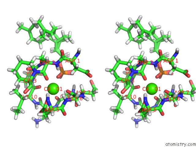
Stereo pair view
Mono view
Stereo pair view
A full contact list of Calcium with other atoms in the Ca binding
site number 1 of Structure of the Laspartomycin C Double Mutant G4D D-Allo-THR9D-Dap in Complex with Geranyl Phosphate within 5.0Å range:
|
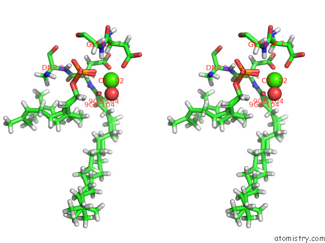
Calcium binding site 2 out of 2 in 7ag5
Go back to
Calcium binding site 2 out
of 2 in the Structure of the Laspartomycin C Double Mutant G4D D-Allo-THR9D-Dap in Complex with Geranyl Phosphate
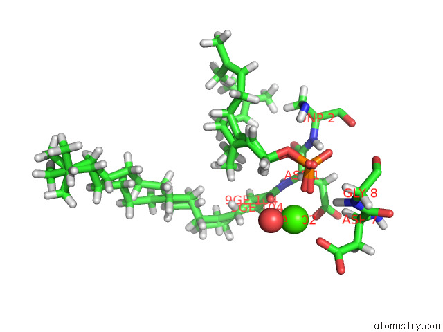
Mono view
Stereo pair view
Mono view
Stereo pair view
A full contact list of Calcium with other atoms in the Ca binding
site number 2 of Structure of the Laspartomycin C Double Mutant G4D D-Allo-THR9D-Dap in Complex with Geranyl Phosphate within 5.0Å range:
|
Reference:
T.M.Wood,
M.R.Zeronian,
N.Buijs,
K.Bertheussen,
H.K.Abedian,
A.V.Johnson,
N.M.Pearce,
M.Lutz,
J.Kemmink,
T.Seirsma,
L.W.Hamoen,
B.J.C.Janssen,
N.I.Martin.
Mechanistic Insights Into the C55-P Targeting Lipopeptide Antibiotics Revealed By Structure-Activity Studies and High-Resolution Crystal Structures Chem Sci V. 13 2985 2022.
ISSN: ESSN 2041-6539
DOI: 10.1039/D1SC07190D
Page generated: Thu Jul 18 22:51:16 2024
ISSN: ESSN 2041-6539
DOI: 10.1039/D1SC07190D
Last articles
Zn in 9MJ5Zn in 9HNW
Zn in 9G0L
Zn in 9FNE
Zn in 9DZN
Zn in 9E0I
Zn in 9D32
Zn in 9DAK
Zn in 8ZXC
Zn in 8ZUF