Calcium »
PDB 7aq2-7b1l »
7ays »
Calcium in PDB 7ays: Structure of Bovine Trypsin Determined From Single Femtosecond Snapshots Per Orientation at Room Temperature
Enzymatic activity of Structure of Bovine Trypsin Determined From Single Femtosecond Snapshots Per Orientation at Room Temperature
All present enzymatic activity of Structure of Bovine Trypsin Determined From Single Femtosecond Snapshots Per Orientation at Room Temperature:
3.4.21.4;
3.4.21.4;
Protein crystallography data
The structure of Structure of Bovine Trypsin Determined From Single Femtosecond Snapshots Per Orientation at Room Temperature, PDB code: 7ays
was solved by
M.Jensen,
with X-Ray Crystallography technique. A brief refinement statistics is given in the table below:
| Resolution Low / High (Å) | 42.53 / 2.10 |
| Space group | P 21 21 21 |
| Cell size a, b, c (Å), α, β, γ (°) | 54.84, 58.6, 67.38, 90, 90, 90 |
| R / Rfree (%) | 21.3 / 26 |
Calcium Binding Sites:
The binding sites of Calcium atom in the Structure of Bovine Trypsin Determined From Single Femtosecond Snapshots Per Orientation at Room Temperature
(pdb code 7ays). This binding sites where shown within
5.0 Angstroms radius around Calcium atom.
In total only one binding site of Calcium was determined in the Structure of Bovine Trypsin Determined From Single Femtosecond Snapshots Per Orientation at Room Temperature, PDB code: 7ays:
In total only one binding site of Calcium was determined in the Structure of Bovine Trypsin Determined From Single Femtosecond Snapshots Per Orientation at Room Temperature, PDB code: 7ays:
Calcium binding site 1 out of 1 in 7ays
Go back to
Calcium binding site 1 out
of 1 in the Structure of Bovine Trypsin Determined From Single Femtosecond Snapshots Per Orientation at Room Temperature
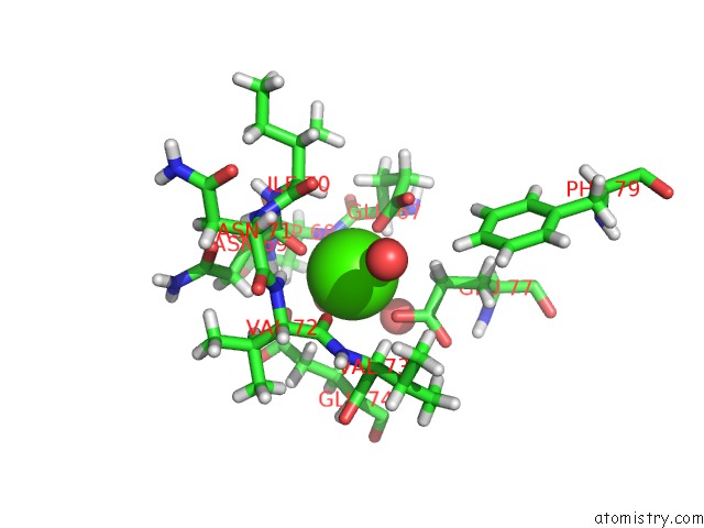
Mono view
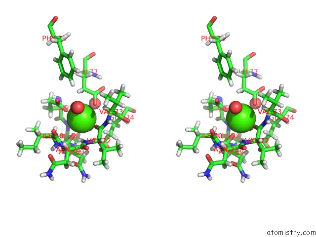
Stereo pair view
Mono view
Stereo pair view
A full contact list of Calcium with other atoms in the Ca binding
site number 1 of Structure of Bovine Trypsin Determined From Single Femtosecond Snapshots Per Orientation at Room Temperature within 5.0Å range:
|
Reference:
M.Jensen,
V.Ahlberg Gagner,
J.Cabello Sanchez,
A.U.J.Bengtsson,
J.C.Ekstrom,
T.Bjorg Ulfarsdottir,
M.J.Garcia-Bonete,
A.Jurgilaitis,
D.Kroon,
V.T.Pham,
S.Checcia,
H.Coudert-Alteirac,
S.Schewa,
M.Rossle,
H.Rodilla,
J.Stake,
V.Zhaunerchyk,
J.Larsson,
G.Katona.
High-Resolution Macromolecular Crystallography at the Femtomax Beamline with Time-Over-Threshold Photon Detection. J.Synchrotron Radiat. V. 28 64 2021.
ISSN: ESSN 1600-5775
PubMed: 33399553
DOI: 10.1107/S1600577520014599
Page generated: Wed Jul 9 20:53:45 2025
ISSN: ESSN 1600-5775
PubMed: 33399553
DOI: 10.1107/S1600577520014599
Last articles
Cd in 2V2LCd in 2R1F
Cd in 2P5Q
Cd in 2V2J
Cd in 2V2I
Cd in 2QTH
Cd in 2QEX
Cd in 2QTF
Cd in 2QA4
Cd in 2QP9