Calcium »
PDB 7b1n-7brw »
7bfz »
Calcium in PDB 7bfz: X-Ray Structure of Human Prostate-Specific Membrane Antigen(Psma) in Complex with A Inhibitor Glu-490
Enzymatic activity of X-Ray Structure of Human Prostate-Specific Membrane Antigen(Psma) in Complex with A Inhibitor Glu-490
All present enzymatic activity of X-Ray Structure of Human Prostate-Specific Membrane Antigen(Psma) in Complex with A Inhibitor Glu-490:
3.4.17.21;
3.4.17.21;
Protein crystallography data
The structure of X-Ray Structure of Human Prostate-Specific Membrane Antigen(Psma) in Complex with A Inhibitor Glu-490, PDB code: 7bfz
was solved by
A.Rakhimbekova,
L.Motlova,
C.Barinka,
with X-Ray Crystallography technique. A brief refinement statistics is given in the table below:
| Resolution Low / High (Å) | 46.96 / 1.73 |
| Space group | I 2 2 2 |
| Cell size a, b, c (Å), α, β, γ (°) | 101.54, 130.103, 158.91, 90, 90, 90 |
| R / Rfree (%) | 16.3 / 18.3 |
Other elements in 7bfz:
The structure of X-Ray Structure of Human Prostate-Specific Membrane Antigen(Psma) in Complex with A Inhibitor Glu-490 also contains other interesting chemical elements:
| Sodium | (Na) | 2 atoms |
| Zinc | (Zn) | 2 atoms |
| Chlorine | (Cl) | 1 atom |
Calcium Binding Sites:
The binding sites of Calcium atom in the X-Ray Structure of Human Prostate-Specific Membrane Antigen(Psma) in Complex with A Inhibitor Glu-490
(pdb code 7bfz). This binding sites where shown within
5.0 Angstroms radius around Calcium atom.
In total only one binding site of Calcium was determined in the X-Ray Structure of Human Prostate-Specific Membrane Antigen(Psma) in Complex with A Inhibitor Glu-490, PDB code: 7bfz:
In total only one binding site of Calcium was determined in the X-Ray Structure of Human Prostate-Specific Membrane Antigen(Psma) in Complex with A Inhibitor Glu-490, PDB code: 7bfz:
Calcium binding site 1 out of 1 in 7bfz
Go back to
Calcium binding site 1 out
of 1 in the X-Ray Structure of Human Prostate-Specific Membrane Antigen(Psma) in Complex with A Inhibitor Glu-490

Mono view
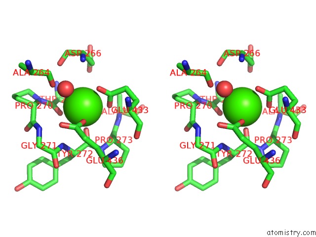
Stereo pair view
Mono view
Stereo pair view
A full contact list of Calcium with other atoms in the Ca binding
site number 1 of X-Ray Structure of Human Prostate-Specific Membrane Antigen(Psma) in Complex with A Inhibitor Glu-490 within 5.0Å range:
|
Reference:
A.Rakhimbekova,
L.Motlova,
C.Barinka.
X-Ray Structure of Human Prostate-Specific Membrane Antigen(Psma) in Complex with A Inhibitor Glu-490 To Be Published.
Page generated: Wed Jul 9 20:59:10 2025
Last articles
Cl in 8BW7Cl in 8BVP
Cl in 8BVS
Cl in 8BUQ
Cl in 8BU5
Cl in 8BU7
Cl in 8BUV
Cl in 8BUC
Cl in 8BSU
Cl in 8BU2